
1 · SNAPSHOT
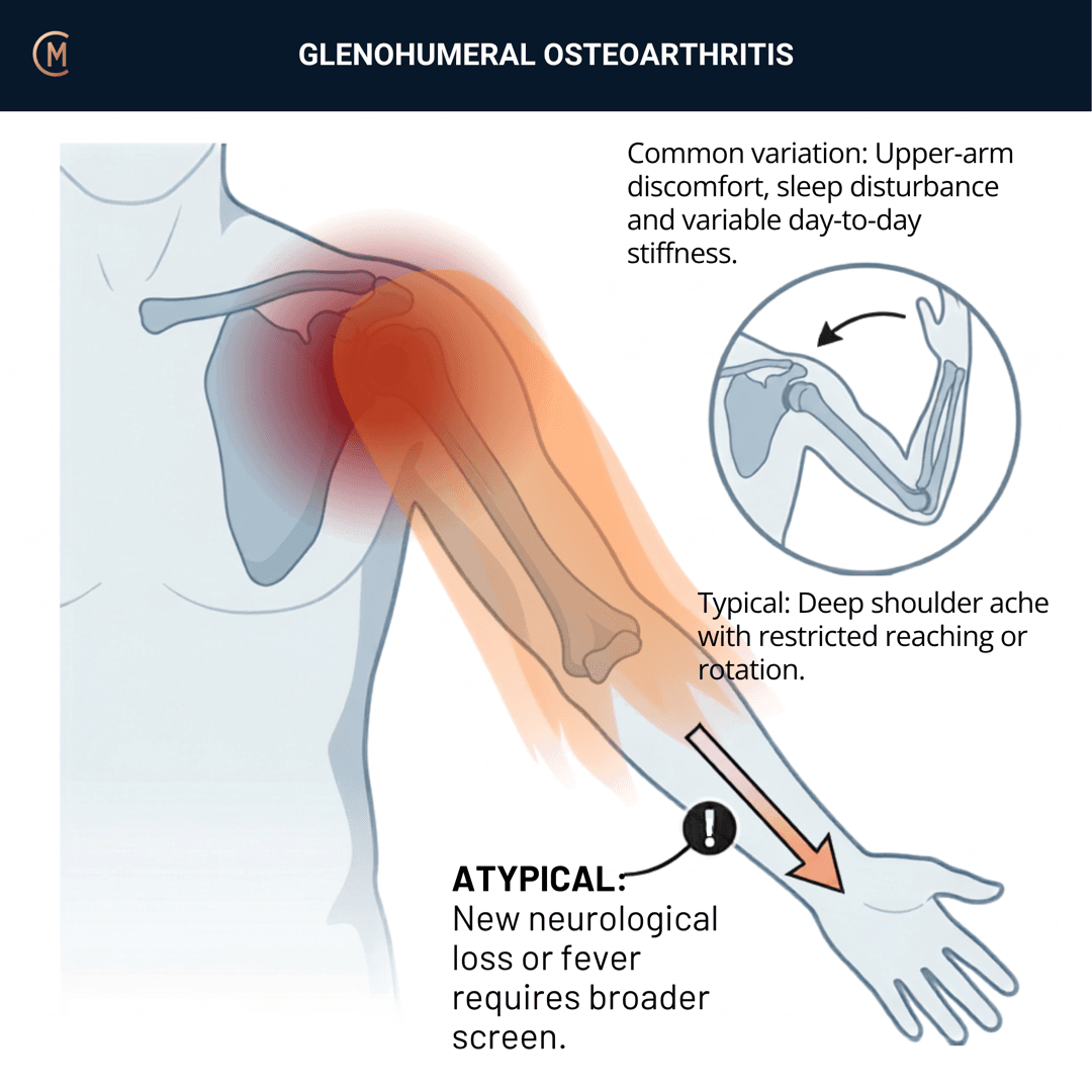
Who: Usually over 50. Also the 30-year-old whose shoulder dislocated at 22.
What: Deep aching pain with progressive stiffness and crepitus. Worse overhead and at end range.
How it behaves: Mechanical and load-related, with morning stiffness that eases as they get moving.
The illness script in one line: a stiff, crepitant, aching shoulder in an older adult — where both active and passive range are lost together, and the plain film will confirm it.
The numbers that matter — and how few of them there are:
| Figure | |
|---|---|
| Share of shoulder complaints | 5–17% |
| Radiographic shoulder OA, middle-aged and elderly | 16–20% |
| Symptomatic GHJ OA 15 years after anterior instability under 40 | 22.7% |
| Australian population prevalence | none exists |
(First two: [1], narrative review, no confidence intervals. Third: [2], n = 154, mean 15.2 years.)
Why those figures are so soft: six competing radiographic classification systems make the true incidence — and therefore the prevalence — unobtainable [3]. And globally, the shoulder isn't even modelled separately: the GBD study reports 595 million people with OA in 2020 with the knee as the most common site, and the shoulder sits inside an unmodelled "other sites" category [4].
Compare that with the knee OA page, where Australia has a national standard and a Cochrane review of 139 trials. Same disease, same joint surface, a fraction of the evidence.
⚠️ No Australian guideline exists for this presentation. Anchored to the APTA Physical Therapist Management of Glenohumeral Joint Osteoarthritis CPG [5], the only physiotherapy-specific guideline in existence, with the AAOS CPG (2020, evidence to June 2019) as the medical and surgical anchor. Australia contributes registry data, not guidance — the AOANJRR.
Pattern > Label. The decision isn't "is it arthritis". It's is this the capsule, the joint surface, or the cuff — and §3 is that decision.
2 · SE — THE SUBJECTIVE PATTERN (the 70%)
Five stages, in your course's order. Insidious, so BEHAVIOUR before HISTORY.
MAIN PROBLEM
The reach they've lost. Seatbelt, back pocket, top shelf, hair, doing up a bra. Same functional list as frozen shoulder — which is exactly why the subjective alone won't separate them.
BODY CHART
- Deep and poorly localised, over the joint line and deltoid. They cup it rather than point.
- May refer to the upper arm. Rarely below the elbow — clear the cervical spine.
BEHAVIOUR
- Aggs: overhead, end range, reaching behind, carrying, lying on it.
- Eases: rest, and — the useful one — movement, after a warm-up period.
- 24-hr pattern: stiff first thing, loosens with movement, sore again by the end of a loaded day.
- Severity: VAS 0–10 at best / at worst / right now.
- Crepitus and grinding the patient volunteers unprompted. Ask, because it's one of the three EULAR-style signs and it's the one that separates this from a capsule.
- Irritability: vigour, severity, time to settle.
HISTORY
-
Gradual, over months to years.
-
The three history features associated with glenohumeral arthritis ([6], systematic review of 21 studies):
Feature Prior shoulder dislocation Age over 75 A diagnosis of knee osteoarthritis The review reports no sensitivities, likelihood ratios or odds ratios in its abstract — so these are associations to ask about, not numbers to quote.
-
Ask about dislocation properly, because this one has figures. In 154 patients under 40 with anterior instability, 22.7% had symptomatic GHJ OA at a mean of 15.2 years [2]. Risk factors within that cohort:
Odds ratio Hyperlaxity 10.1 (95% CI 1.4–72.4) Manual labourer 6.1 (1.02–36.1) Smoking 4.3 (1.1–16.5) Wide intervals, small numbers — but a 25-year-old with a dislocated shoulder is someone whose future you can actually mention.
-
Previous imaging, and what they were told. See §5.
SPECIAL / SCREENING QUESTIONS
General health, weight loss, fever, malignancy history, inflammatory features, medications, neurological symptoms. See §4.
A note on how thin this is: the evidence base for this pattern holds almost no subjective research — most of it is surgical. The best published description of the presenting pattern is one sentence in a family-medicine review: over 50, it "usually presents as gradual pain and loss of motion" [7]. That is the state of the literature.
Assessing this region? Download the Shoulder assessment template →
3 · HYPOTHESIS RANKING
| Competing pattern | The one discriminating feature |
|---|---|
| Adhesive capsulitis | Passive external rotation near-obliterated, firm end-feel, no crepitus. Pain came before stiffness |
| Massive rotator cuff tear | Active elevation lost, passive preserved. Pseudoparalysis, scapular hitch |
| Rotator cuff-related pain | Painful arc with near-full passive range in every direction |
| Calcific tendinopathy | Sudden, severe, guarded. Pain-limited, not truly restricted |
| Not the shoulder | Doesn't behave mechanically, or symptoms below the elbow → §4 |
The discrimination, and the honest state of it
This page's central problem: a capsular pattern (ER > ABD > F) is commonly taught for GHJ OA, and there is no evidence for it.
The only study that directly tested whether a consistent capsular pattern exists in stiff shoulders concluded, in its own words: "No single capsular pattern emerged" [8]. The Cyriax capsular pattern is a teaching convention. No study has measured the presenting range profile in GHJ OA at all.
What that same study does give you is the one measured anchor in this whole comparison — and it belongs to the other condition:
| In frozen shoulder (n = 10) | Mean peak humerothoracic motion |
|---|---|
| External rotation at the side | 4.5° |
| ER in abduction | 33.5° |
| Abduction | 98.4° |
| Flexion | 116.9° |
So teach magnitude, not pattern. External rotation that has collapsed to near-nothing with a firm end-feel and no crepitus is a capsule. External rotation reduced but present, with a hard end-feel and crepitus, is a joint surface. Active loss with preserved passive is a cuff.
And say the second part out loud: that contrast is reasoning, not a validated rule. One of those three descriptions has a number behind it. The other two do not.
Jones' hypothesis categories that change your plan: sources of symptoms (capsule vs joint surface vs cuff), pathophysiology (this one is genuinely structural, unlike most of what you'll see), and prognosis.
Same stiff shoulder, three stories. Crepitus and a hard end-feel is the joint. No crepitus and a firm end-feel is the capsule. Active-only loss is the cuff.
4 · RED FLAGS — DON'T MISS THIS
This section is free, on every pattern, forever.
The honest position
Primary-care red-flag evidence for this pattern is thin — most of the safety literature is post-arthroplasty complication reviews. The screen below comes from a primary-care review with Australian senior authorship [9].
What to actually screen
| Signs / symptoms | Implication |
|---|---|
| Fever, unexplained weight loss, malignancy history | Infection · malignant neoplasm — and the trigger for early imaging |
| Pain not behaving mechanically, unchanged by arm position | Non-shoulder referred pain |
| Significant functional or neurological deficit | Nerve injury · cervical radiculopathy · massive tear |
| Systemic inflammatory features, multiple joints, prolonged morning stiffness | Inflammatory arthropathy |
| Trauma with inability to elevate, or sudden deformity | Fracture · dislocation · acute tear |
| Persistent or worsening pain and debility despite appropriate care | Re-examine the diagnosis, then refer |
Act on
Early imaging is not indicated in the absence of significant trauma or a suspicious feature. That is the review's position, and it is worth holding alongside §5 — where the film is genuinely the answer once you have decided to ask the question.
If they've had a shoulder replacement, the red flags change. Sudden focal pain with functional loss after a reverse arthroplasty can be an acromial or scapular spine fracture, and plain films can miss it — that needs a CT [20]. Instability and periprosthetic fracture are the two complications to know by name.
Fever, weight loss, cancer history — that is when you image early, not when the shoulder is stiff.
Pain that doesn't change with arm position isn't the shoulder.
A post-arthroplasty shoulder with sudden focal pain needs more than an X-ray.
5 · OE — THE OBJECTIVE (the 30%)
Members
Sections 5 to 7 are for members
The objective exam, management and pearls, plus the six-minute lesson and the debrief. Free with an account.
Sign in or join free6 · MX — MANAGEMENT ESSENTIALS
Members
Sections 5 to 7 are for members
The objective exam, management and pearls, plus the six-minute lesson and the debrief. Free with an account.
Sign in or join free7 · CLINICAL PEARLS + RETRIEVAL CHECK
Members
Sections 5 to 7 are for members
The objective exam, management and pearls, plus the six-minute lesson and the debrief. Free with an account.
Sign in or join freeMembers
The 4-minute lesson is for members
Twelve slides on this pattern: what you'll hear, what to examine, what to do. Free with an account.
Sign in or join freeGuideline anchor
No Australian guideline for this presentation.
More
Anchored to Physical Therapist Management of Glenohumeral Joint Osteoarthritis — APTA Clinical Practice Guideline (Michener et al.)
No Australian guideline or clinical care standard exists for glenohumeral OA. The ACSQHC standard covers the knee only; the RACGP guideline covers knee and hip only. Australia does have registry data — the AOANJRR.
Members
The evidence summary is for members
Verified diagnostic accuracy, where the evidence stands and what it doesn't settle. Free with an account.
Sign in or join freeSources · 19
ShowHide
Every source on this page resolves and title-verifies against PubMed. Newest first.
- 2021Ibounig et al. (2021). Glenohumeral osteoarthritis: an overview of etiology and diagnostics. Scandinavian journal of surgery : SJS : official organ for the Finnish Surgical Society and the Scandinavian Surgical Society, 110(3), 441-451. https://doi.org/10.1177/1457496920935018
- 2020Kruckeberg et al. (2020). Incidence of and Risk Factors for Glenohumeral Osteoarthritis After Anterior Shoulder Instability: A US Population-Based Study With Average 15-Year Follow-up. Orthopaedic journal of sports medicine, 8(11), 2325967120962515. https://doi.org/10.1177/2325967120962515
- 2024Crane et al. (2024). Glenohumeral Osteoarthritis: A Biological Advantage or a Missed Diagnosis?. Journal of clinical medicine, 13(8). https://doi.org/10.3390/jcm13082341
- 2023Anon. (2023). Global, regional, and national burden of osteoarthritis, 1990-2020 and projections to 2050: a systematic analysis for the Global Burden of Disease Study 2021. The Lancet. Rheumatology, 5(9), e508-e522. https://doi.org/10.1016/S2665-9913(23)00163-7
- 2023Michener et al. (2023). Physical Therapist Management of Glenohumeral Joint Osteoarthritis: A Clinical Practice Guideline from the American Physical Therapy Association. Physical therapy, 103(6). https://doi.org/10.1093/ptj/pzad041
- 2016Raynor & Kuhn (2016). Utility of features of the patient's history in the diagnosis of atraumatic shoulder pain: a systematic review. Journal of shoulder and elbow surgery, 25(4), 688-94. https://doi.org/10.1016/j.jse.2015.09.023
- 2008Burbank et al. (2008). Chronic shoulder pain: part I. Evaluation and diagnosis. American family physician, 77(4), 453-60. PubMed 18326164
- 2003Rundquist et al. (2003). Shoulder kinematics in subjects with frozen shoulder. Archives of physical medicine and rehabilitation, 84(10), 1473-9. https://doi.org/10.1016/s0003-9993(03)00359-9
- 2026Haas et al. (2026). Management of Shoulder Pain in Primary Care: A Review. JAMA internal medicine. https://doi.org/10.1001/jamainternmed.2026.3135
- 2008Jia et al. (2008). Clinical evaluation of the shoulder shrug sign. Clinical orthopaedics and related research, 466(11), 2813-9. https://doi.org/10.1007/s11999-008-0331-3
- 2014Matsen FA 3rd & Gupta (2014). Axillary view: arthritic glenohumeral anatomy and changes after ream and run. Clinical orthopaedics and related research, 472(3), 894-902. https://doi.org/10.1007/s11999-013-3327-6
- 2024Ibounig et al. (2024). Systematic Review of Shoulder Imaging Abnormalities in Asymptomatic Adult Shoulders (SCRUTINY): Abnormalities of the glenohumeral joint. Osteoarthritis and cartilage, 32(10), 1184-1196. https://doi.org/10.1016/j.joca.2024.06.001
- 2023Razmjou et al. (2023). Assessing Appropriateness for Shoulder Arthroplasty Using a Shared Decision-Making Process. Journal of shoulder and elbow arthroplasty, 7, 24715492231167104. https://doi.org/10.1177/24715492231167104
- 2017Macías-Hernández et al. (2017). Glenohumeral osteoarthritis: overview, therapy, and rehabilitation. Disability and rehabilitation, 39(16), 1674-1682. https://doi.org/10.1080/09638288.2016.1207206
- 2026Gayretli Atan et al. (2026). Are manual therapy applications an effective treatment approach in patients with glenohumeral arthritis? A randomized controlled trial. Journal of hand therapy : official journal of the American Society of Hand Therapists, 39(3), 646-655. https://doi.org/10.1016/j.jht.2025.12.016
- 2025Migliorini et al. (2025). Intra-articular injections for shoulder arthritis in adults: a systematic review. European journal of medical research, 30(1), 1080. https://doi.org/10.1186/s40001-025-03423-4
- 2022Beck Larsen et al. (2022). Study protocol: Prosthesis versus Active (ProAct) exercise program in patients with glenohumeral osteoarthritis - a multicenter, randomized controlled trial. Acta orthopaedica, 93, 303-316. https://doi.org/10.2340/17453674.2022.2043
- 2023Khoriati et al. (2023). Survivorship of shoulder arthroplasty in young patients with osteoarthritis: an analysis of the Australian Orthopaedic Association National Joint Replacement Registry. Journal of shoulder and elbow surgery, 32(10), 2105-2114. https://doi.org/10.1016/j.jse.2023.03.024
- 2021Ravi et al. (2021). Outcome and complications following revision shoulder arthroplasty : a systematic review and meta-analysis. Bone & joint open, 2(8), 618-630. https://doi.org/10.1302/2633-1462.28.BJO-2021-0092.R1
Mapped to this pattern
Generic measures that also apply
- Borg RPE
Borg Rating of Perceived Exertion Scale (6-20)
perceived exertion · 1 items · 1 min
- GROC
Global Rating of Change Scale
perceived change since baseline · 1 items · 1 min
- NPRS
Numeric Pain Rating Scale
pain intensity · 1 items · 1 min
- PGIC
Patient Global Impression of Change
perceived change since baseline · 1 items · 1 min
- PSFS
Patient-Specific Functional Scale
patient-generated function · 3 items · 2 min
- SANE
Single Assessment Numeric Evaluation
patient-rated function as a percentage of normal · 1 items · 1 min
- VAS
Visual Analogue Scale
pain intensity · 1 items · 1 min
- Borg CR10
Borg Category Ratio 10 Scale
perceived exertion or symptom intensity, ratio scale · 1 items · 1 min
- GAS
Goal Attainment Scaling
individualised goal achievement · 10 min
Did this page do its job?
CliniStudy is in beta. One tap is enough — it decides what gets fixed first.