
1 · SNAPSHOT
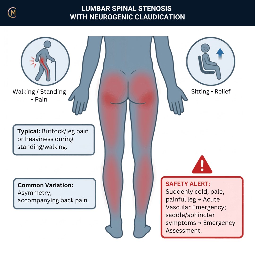
Who: Typically over 60. Gradual onset over months to years, fluctuating rather than linear.
What: Back and leg pain, heaviness or "tired legs", often bilateral, with a walking distance they can quote you in metres.
How it behaves: Aggs: walking, standing, downhill walking, lumbar extension. Eases: sitting, bending forward, leaning on a trolley, cycling, walking uphill.
The illness script in one line:
Extension closes the canal and walking loads it — so the legs give out at a repeatable distance, and the position that fixes it is flexion, not rest.
The one thing to take from this page:
"Stenosis" means three different things and the literature swaps between them mid-sentence. A radiological finding. A clinical syndrome. A diagnosis. You assess the second one.
Say what you mean by the word, before you say anything else
| What it is | Who owns it | |
|---|---|---|
| 1 · The finding | Narrowing of the central canal, lateral recess or foramen on CT or MRI. Common, age-related and frequently silent. | The radiologist |
| 2 · The syndrome | Neurogenic claudication — buttock and leg pain or heaviness on walking and standing, eased by sitting or flexion, in an older adult. | You. This is the whole page. |
| 3 · The diagnosis | The syndrome plus imaging that plausibly explains it, minus the things that mimic it. | The medical team, with your reasoning in the referral |
Hold that line all the way down the page. The pattern on this page describes number 2. Most of the imaging evidence describes number 1. They overlap far less than students expect, and that gap is the most useful thing here.
| Figure | Number |
|---|---|
| Community adults with more than moderate central stenosis on MRI | 77.9% |
| Same cohort with severe central stenosis | 30.4% |
| Of that severe group, how many were symptomatic | 17.5% |
| Acquired relative stenosis on CT, under 40 yrs → 60–69 yrs | 20.0% → 47.2% |
| The syndrome in a population-based cohort | 9.3% (95% CI 7.7–11.3) |
| Clinical prevalence, general population / primary care | 11% (4–18%) / 25% (19–32%) |
| MRI severity vs pain, walking capacity or disability | no correlation with any parameter |
| Peripheral arterial disease in confirmed stenosis | 6.7% — and half of those were undetected |
| Screen-positive community dwellers who were negative a year later | 54% |
| Natural course to 3 years without surgery | ~⅓ improve · ~50% unchanged · 10–20% worse |
[1][2][3][4][5][6][7][8]
There is no Australian guideline for this pattern, and that is not a formatting note. The ACSQHC Low Back Pain Clinical Care Standard (2022) was read in full on 2 Sep 2026. The words "stenosis", "claudication" and "neurogenic" appear zero times — not in the eight quality statements, not in the clinician sections, not in the consumer sections. It covers "low back pain, with or without leg pain or other neurological symptoms", it names sciatica and radicular pain, and it lists its differentials as nephritis, colitis, hip osteoarthritis and aortic dissection. Stenosis is not one of them.
So split it honestly. The generic layer — assessment, psychosocial screening, when not to image, when to escalate — does sit under an Australian standard, and you should say so. The pattern-specific layer — the claudication history, the vascular differential, the walking programme, the drug list, the surgical threshold — is anchored to Bussières et al. 2021 [9], a Canadian-led international GRADE guideline. Not Australian. When your educator asks "says who?", name which half you are quoting.
Patterns > Labels. "L4/5 canal stenosis" names a measurement. Older adult, bilateral leg heaviness, walking limit in metres, eased by flexion, vessels checked names the problem you can manage this week — and it is the one you take to your educator.
2 · SE — THE SUBJECTIVE PATTERN (the 70%)
Five stages, in order. On this pattern the history is not the lead-in to the diagnosis — it is the diagnostic instrument, and that is not a slogan, it is the accuracy data. A self-administered history questionnaire reached sensitivity 84% / specificity 78% in a 250-patient validation set [10]. Head to head in 3,331 consecutive low back pain patients, of whom 42.5% had stenosis, the LSS diagnosis support tool hit 91.3% / 76.0% against the NASS clinical description at 63.9% / 89.5% [11]. Nothing in section 5 comes close to those numbers.
MAIN PROBLEM
"I can't walk to the shops any more. About 200 metres and both legs go heavy and dead, and I have to sit down. Sitting fixes it in a couple of minutes. Then I can go again."
Listen for a distance and a position. The main problem in this pattern is almost never "my back hurts" — it is a walking limit and a posture that resets it. If they lead with a number in metres, you are already most of the way there.
BODY CHART
- Map P1 and P2 bilaterally. Stenosis usually gives you two columns on the body chart, not one — buttock, posterior thigh, calf, often both sides, often asymmetrically.
- Mark heaviness, deadness and "tired legs" as distinct from pain. Patients under-report these because they don't sound like a symptom.
- Mark paraesthesia and numbness separately, and mark the perineum — see section 4 before you write anything in that box.
- Back pain is often the smaller half. Leg-dominant, below the buttock, bilateral is the shape.
- Draw the other leg even when they say it's fine. Bilateral is the pattern; it is also the CES question.
BEHAVIOUR
This is where the diagnosis lives, so code it properly.
- Coding: In — intermittent, activity-dependent, position-driven. A constant (P) presentation should make you re-ask the question. Morning stiffness here is usually M (<30 min).
- Aggs: walking, standing still, downhill walking, lumbar extension, reaching up, long queues.
Eases: sitting, bending forward, leaning on a trolley or a bench, cycling, walking uphill, going up stairs more easily than down. - 24-hr pattern: walking-dose dependent, not clock dependent — walking distance to onset is the number.
- Night: sitting and bending forward are the Eases. Night rest pain is in the vascular screen in §4 — ask it.
- Severity: VAS 0–10 at best / at worst / right now.
- Three numbers, and they are your asterisk signs and your outcome measure:
- Walking distance to onset — in metres or minutes, not "a while".
- The position that relieves it — sitting, or flexion, or does it need both.
- Time to settle once they adopt it.
- Irritability, all three parts — resting symptoms + vigour of the provoking activity + time to settle. Here it sets your walking dose, not your technique choice.
- Ask the four Nadeau items explicitly, in these words: shopping trolley sign · symptoms above or below the knee · triggered by standing alone · relieved by sitting. All four present → +LR 13 for neurogenic. And the single most useful negative you own: no pain triggered by standing alone → −LR 0.05 ([12], n=53, no published confidence intervals).
HISTORY
Give this stage the time it deserves. On this pattern it carries more diagnostic weight than anything you will do with your hands.
- Onset gradual, over months to years. A sudden onset is a different pattern until proven otherwise.
- The trajectory, honestly asked. Getting worse, or fluctuating? Most patients say worse. Most cohorts say fluctuating — and 54% of community screen-positives were screen-negative a year later [7]. Ask them what a good month looked like.
- The walking limit over time. Was it 2 km last year and 200 m now, or has it been 200 m for five years? Rate of change is the referral question, not the distance itself.
- Konno's five history items are the fastest screen you have — each carries an OR ≥2, p<0.05 for the syndrome:
| The five items | |
|---|---|
| Age >50 | ✔ / ✘ |
| Lower-limb pain or numbness | ✔ / ✘ |
| ↑ pain when walking | ✔ / ✘ |
| ↑ pain when standing | ✔ / ✘ |
| Relief on bending forward | ✔ / ✘ |
- Vascular history, taken properly, not ticked. Smoking, diabetes, ischaemic heart disease, previous stroke, hypertension, cholesterol, claudication in the family. This is a screening question in every other lumbar pattern. Here it is part of the differential (section 4).
- Previous imaging, and — more importantly — what they were told about it. Someone managing "severe spinal stenosis" in their head is managing a different problem from the one in their legs.
- Comorbid knee and hip OA. Self-reported stenosis symptoms are reported by 8–50% of primary-care knee and hip OA patients, and by 11–71% of the low back pain cohort — yet fewer than 10% of each met clinical stenosis criteria ([13], n=10,234). Symptoms are not the syndrome.
SPECIAL / SCREENING QUESTIONS
Every patient. Every time. Before you touch them. This population is older, so the base rates of the dangerous things are higher than in a young back — see section 4 for the numbers, and go there before you go to section 5.
- Cauda equina, asked directly and unhurriedly: bladder, bowel, saddle or perineal sensory change, sexual dysfunction, bilateral progressive weakness.
- Vascular claudication and PAD: calf symptoms, cold feet, non-healing wounds, rest pain at night, smoking.
- Abdominal aortic aneurysm: non-mechanical abdominal or back pain that does not answer to position.
- Malignancy, fracture, infection, inflammatory — carry over unchanged from the NSLBP page. Screen them; don't learn them twice.
- Medications, falls history, and what they've stopped doing. Sexual function and social isolation belong in this stage too — pain plus limited walking drives hopelessness, anxiety and isolation, and no guideline prompts you to ask ([14]; narrative review, framing only).
CLOSE THE SUBJECTIVE PROPERLY
Before you stand up you should be able to say, out loud: the distance, the relieving position, the settle time, whether the four Nadeau items are present, and whether the calf-plus-standing vascular pattern is there instead. That sentence is your reasoning. The objective exists to test it.
Assessing this region? Download the Lumbar Spine & SIJ assessment template →
3 · HYPOTHESIS RANKING
Top three, one discriminating feature each. Rule out the two dangerous ones first, then split the two that actually look alike.
| Rank | Pattern | The one discriminating feature |
|---|---|---|
| 1 | Cauda equina syndrome | Bladder or bowel change, saddle or perineal sensory loss, bilateral progressive weakness, sexual dysfunction. Present → same-day emergency referral, no exceptions. The trap: perineal numbness also raises the posttest probability of stenosis by >25% [15]. It belongs to both lists. CES is excluded first, regardless. See the cauda equina syndrome page — it owns this content; don't rely on this row. |
| 2 | Vascular claudication / peripheral arterial disease | Calf symptoms relieved by standing alone, without needing to sit or lean forward: +LR 20.0. Neurogenic needs the flexed position, not just the stop. Cold foot, absent pedal pulses, hair loss, non-healing ulcer, smoker, diabetic. If the picture is atypical, the ankle-brachial index does the discriminating — sensitivity 85.3%, specificity 85.7% [12][16]. And they coexist in 6.7%. Neither excludes the other [6]. |
| 3 | Lumbar radicular pain / radiculopathy | Leg pain worse than back pain, below the knee, in a dermatomal band, usually unilateral, worse with sitting, coughing and flexion, eased by standing. That is the mirror image of the stenosis behaviour. Stenosis is usually bilateral, extension-provoked and flexion-eased. See the lumbar radicular pain page. |
And three more you should be able to name:
- Abdominal aortic aneurysm — same demographic, same risk factors. The discriminator is positional dependence: stenosis pain answers to flexion and to stopping; an aneurysm does not. Non-mechanical back or abdominal pain, a pulsatile mass, pain unrelated to walking distance → same-day medical assessment. Sudden severe or tearing pain, collapse, or a patient who looks shocked → ambulance now. This page carries no accuracy data for AAA and will not pretend to. It is a screening obligation, not a test.
- Lumbar discogenic pain — central or bilateral back pain with non-dermatomal buttock and thigh referral, dull and deep, worse with sitting and flexion, eased by standing and walking. Flexion is the aggravator, not the reliever. No walking-distance limit.
- Hip osteoarthritis — groin pain, pain on weight-bearing from the first step rather than after a distance, restricted and painful hip IR, and no relief from sitting because the problem is not in the canal. Named as a differential by the ACSQHC standard itself, common in the same age group, and it coexists constantly.
The honest bit. Nobody has measured how reliably two clinicians agree on this diagnosis. There is no κ study for lumbar spinal canal stenosis the way there is for sciatica. We know the questionnaires perform decently against a physician's diagnosis; we do not know how well two physiotherapists agree with each other. Take your ranking to your educator with the discriminator attached — "bilateral, 200 m limit, eased by flexion, calf symptoms need sitting not just standing, pedal pulses present, so I've ranked it neurogenic" is a reasoning conversation. "I think it's stenosis" isn't.
4 · RED FLAGS — DON'T MISS THIS
This section is free, on every pattern, forever. Safety is never gated.
Start here, and start with the reason this page's red flags differ from a young back's: the population is older. Same questions, different base rates — and a base rate is what turns a screening question into a finding.
| The dangerous thing | What age does to the odds |
|---|---|
| Peripheral arterial disease, high-income countries | 3.54% (95% CI 1.17–10.24) at 40–44 yrs → 21.24% (15.22–28.90) at 80–84 yrs |
| Spinal fracture, posttest probability from older age alone | 9% (95% CI 3–25%) |
| Spinal cancer, posttest probability from history of malignancy | 33% (22–46%) — the highest single flag there is |
| Fracture, when multiple red flags are present together | 90% (34–99%) |
| Cauda equina as a share of primary-care low back pain | 0.08% — still rare, still the emergency |
([17], 118 articles [18], 14 studies, 53 red flags — this review carries an erratum that has not been read, so only the abstract's posttest probabilities appear here [19].)
Count them, don't tick them. Downie's finding is that most red flags in current guidelines change probability virtually not at all on their own — and that several together change it enormously. One flag is a question. Three flags is a phone call.
Cauda equina syndrome — and the perineal numbness trap
This is the one place on the page where a student can genuinely take the wrong turn, so read it slowly.
Perineal numbness is a cardinal cauda equina symptom. It is also one of the six findings that raise the posttest probability of lumbar spinal canal stenosis by more than 25% [15]. The same symptom sits on both lists, pointing at an emergency on one and at your working pattern on the other.
Which way do you jump? You don't. There is no branch here.
CES is excluded first, regardless. Perineal numbness never gets filed under "that's just the stenosis" — not because stenosis can't cause it, but because you cannot tell the difference in the room, and only one of the two answers is time-critical.
Ask, in plain language, every visit:
- Any change in bladder? Retention, poor stream, reduced sensation of filling, leaking without knowing?
- Any change in bowel control?
- Any numbness in the saddle or perineum — wiping, sitting, sex?
- Are both legs getting weaker, and is it progressing?
Any positive → same-day emergency referral. Do not finish your P/E. Do not book a review. And a negative screen is not a discharge — the cauda equina syndrome page carries the accuracy data, the base rates and the referral pathway in full. It owns that content. Go and read it there; it is free too.
Vascular claudication and peripheral arterial disease
"Possible vascular comorbidities" is how it is often put. That is under-stated, and this is the correction.
- 38 of 570 consecutive imaging-confirmed stenosis patients across 64 facilities had PAD — 6.7%. Twenty (3.5%) already knew. Eighteen (3.2%) did not — half the total, undetected at enrolment [6].
- And the coexisting group did worse. At three months, the PAD group improved less in buttock and leg pain and in SF-36 general health (both p<0.05).
- So: finding stenosis does not clear the vessels. They are not alternatives; they are neighbours in the same demographic.
Screen it physically, every time. Pedal pulses (dorsalis pedis, posterior tibial), foot temperature and colour, capillary refill, hair loss, non-healing wounds. Ask about smoking, diabetes, ischaemic heart disease, previous stroke and night rest pain.
Atypical picture → medical referral for an ankle-brachial index. The discriminating numbers are in section 5, and so is the reason you must not read an ABI naively in a diabetic patient.
Abdominal aortic aneurysm
Same demographic, same risk factors, and it does not care about your pattern. The discriminator is positional dependence — stenosis pain answers to flexion and to stopping; an aneurysm does not. Non-mechanical back or abdominal pain unrelated to walking distance, or a pulsatile abdominal mass, in a patient who is otherwise well → same-day medical assessment. Sudden severe or tearing back or abdominal pain, collapse, sweating or a patient who looks shocked → ambulance now — possible rupture. See the abdominal aortic aneurysm page. This page carries no accuracy data for AAA. It is a screening obligation, not a test you perform.
The rest of the lumbar flags
Malignancy, fracture, infection, inflammatory — the questions themselves are unchanged and live on the NSLBP page. Screen them here with the older-adult base rates above in your head; don't learn them twice.
Bladder + saddle = surgical emergency.
Calf + standing-relieved = check the arteries, not the canal.
If the walking limit is falling week to week, or the weakness is progressing → refer, don't rehab.
5 · OE — THE OBJECTIVE (the 30%)
Members
Sections 5 to 7 are for members
The objective exam, management and pearls, plus the six-minute lesson and the debrief. Free with an account.
Sign in or join free6 · MX — MANAGEMENT ESSENTIALS
Members
Sections 5 to 7 are for members
The objective exam, management and pearls, plus the six-minute lesson and the debrief. Free with an account.
Sign in or join free7 · CLINICAL PEARLS + RETRIEVAL CHECK
Members
Sections 5 to 7 are for members
The objective exam, management and pearls, plus the six-minute lesson and the debrief. Free with an account.
Sign in or join freeMembers
The 4-minute lesson is for members
Twelve slides on this pattern: what you'll hear, what to examine, what to do. Free with an account.
Sign in or join freeGuideline anchor
No Australian guideline for this presentation. Anchored to Non-surgical interventions for lumbar spinal stenosis leading to neurogenic claudication: a clinical practice guideline (Bussières et al. 2021, J Pain, PMID 33857615, GRADE).
More
Supplemented by the updated systematic review it now rests on (Ammendolia et al. 2022, PMID 35046008). This is a CANADIAN-LED INTERNATIONAL guideline, not an Australian one, and the page says so in the banner.
No Australian guideline or clinical care standard covers lumbar spinal canal stenosis. Verified live against safetyandquality.gov.au on 2 Sep 2026: the ACSQHC Low Back Pain Clinical Care Standard (2022) contains the words 'stenosis', 'claudication' and 'neurogenic' ZERO times across its eight quality statements and its clinician and consumer sections. It covers 'low back pain, with or without leg pain or other neurological symptoms', names sciatica and radicular pain, and lists its differentials as nephritis, colitis, hip osteoarthritis and aortic dissection. Stenosis is not one of them. So the generic assessment, psychosocial screening, imaging and escalation layer of this page DOES sit under an Australian standard; the pattern-specific care does not.
Members
The evidence summary is for members
Verified diagnostic accuracy, where the evidence stands and what it doesn't settle. Free with an account.
Sign in or join freeSources · 32
ShowHide
Every source on this page resolves and title-verifies against PubMed. Newest first.
- 2013Ishimoto et al. (2013). Associations between radiographic lumbar spinal stenosis and clinical symptoms in the general population: the Wakayama Spine Study. Osteoarthritis and cartilage, 21(6), 783-8. https://doi.org/10.1016/j.joca.2013.02.656
- 2009Kalichman et al. (2009). Spinal stenosis prevalence and association with symptoms: the Framingham Study. The spine journal : official journal of the North American Spine Society, 9(7), 545-50. https://doi.org/10.1016/j.spinee.2009.03.005
- 2012Ishimoto et al. (2012). Prevalence of symptomatic lumbar spinal stenosis and its association with physical performance in a population-based cohort in Japan: the Wakayama Spine Study. Osteoarthritis and cartilage, 20(10), 1103-8. https://doi.org/10.1016/j.joca.2012.06.018
- 2020Jensen et al. (2020). Prevalence of lumbar spinal stenosis in general and clinical populations: a systematic review and meta-analysis. European spine journal : official publication of the European Spine Society, the European Spinal Deformity Society, and the European Section of the Cervical Spine Research Society, 29(9), 2143-2163. https://doi.org/10.1007/s00586-020-06339-1
- 2018Andrasinova et al. (2018). Is there a Correlation Between Degree of Radiologic Lumbar Spinal Stenosis and its Clinical Manifestation?. Clinical spine surgery, 31(8), E403-E408. https://doi.org/10.1097/BSD.0000000000000681
- 2012Uesugi et al. (2012). Lumbar spinal stenosis associated with peripheral arterial disease: a prospective multicenter observational study. Journal of orthopaedic science : official journal of the Japanese Orthopaedic Association, 17(6), 673-81. https://doi.org/10.1007/s00776-012-0311-z
- 2018Otani et al. (2018). Prospective one-year follow-up of lumbar spinal stenosis in a regional community. Journal of pain research, 11, 455-464. https://doi.org/10.2147/JPR.S148402
- 2022Katz et al. (2022). Diagnosis and Management of Lumbar Spinal Stenosis: A Review. JAMA, 327(17), 1688-1699. https://doi.org/10.1001/jama.2022.5921
- 2021Bussières et al. (2021). Non-Surgical Interventions for Lumbar Spinal Stenosis Leading To Neurogenic Claudication: A Clinical Practice Guideline. The journal of pain, 22(9), 1015-1039. https://doi.org/10.1016/j.jpain.2021.03.147
- 2007Konno et al. (2007). A diagnostic support tool for lumbar spinal stenosis: a self-administered, self-reported history questionnaire. BMC musculoskeletal disorders, 8, 102. https://doi.org/10.1186/1471-2474-8-102
- 2022Tominaga et al. (2022). Diagnostic accuracy of the lumbar spinal stenosis-diagnosis support tool and the lumbar spinal stenosis-self-administered, self-reported history questionnaire. PloS one, 17(5), e0267892. https://doi.org/10.1371/journal.pone.0267892
- 2013Nadeau et al. (2013). The reliability of differentiating neurogenic claudication from vascular claudication based on symptomatic presentation. Canadian journal of surgery. Journal canadien de chirurgie, 56(6), 372-7. https://doi.org/10.1503/cjs.016512
- 2021Young et al. (2021). Symptoms of lumbar spinal stenosis in people with knee or hip osteoarthritis or low back pain: a cross-sectional study of 10,234 participants in primary care. Osteoarthritis and cartilage, 29(11), 1515-1520. https://doi.org/10.1016/j.joca.2021.07.012
- 2025Ammendolia (2025). No drugs, more sex? And Rock'n Roll: effective non-operative treatments and practical management strategies for older adults with lumbar spinal stenosis. Chiropractic & manual therapies, 33(1), 27. https://doi.org/10.1186/s12998-025-00590-3
- 2020Cook et al. (2020). Systematic review of diagnostic accuracy of patient history, clinical findings, and physical tests in the diagnosis of lumbar spinal stenosis. European spine journal : official publication of the European Spine Society, the European Spinal Deformity Society, and the European Section of the Cervical Spine Research Society, 29(1), 93-112. https://doi.org/10.1007/s00586-019-06048-4
- 2012Jeon et al. (2012). The validity of ankle-brachial index for the differential diagnosis of peripheral arterial disease and lumbar spinal stenosis in patients with atypical claudication. European spine journal : official publication of the European Spine Society, the European Spinal Deformity Society, and the European Section of the Cervical Spine Research Society, 21(6), 1165-70. https://doi.org/10.1007/s00586-011-2072-3
- 2019Song et al. (2019). Global, regional, and national prevalence and risk factors for peripheral artery disease in 2015: an updated systematic review and analysis. The Lancet. Global health, 7(8), e1020-e1030. https://doi.org/10.1016/S2214-109X(19)30255-4
- 2013Downie et al. (2013). Red flags to screen for malignancy and fracture in patients with low back pain: systematic review. BMJ (Clinical research ed.), 347, f7095. https://doi.org/10.1136/bmj.f7095
- 2020Hoeritzauer et al. (2020). What is the incidence of cauda equina syndrome? A systematic review. Journal of neurosurgery. Spine, 32(6), 832-841. https://doi.org/10.3171/2019.12.SPINE19839
- 2021Houle et al. (2021). Comparison of walking variations during treadmill walking test between neurogenic and vascular claudication: a crossover study. Chiropractic & manual therapies, 29(1), 24. https://doi.org/10.1186/s12998-021-00382-5
- 1997Fritz et al. (1997). Preliminary results of the use of a two-stage treadmill test as a clinical diagnostic tool in the differential diagnosis of lumbar spinal stenosis. Journal of spinal disorders, 10(5), 410-6. PubMed 9355058
- 2013Minamide et al. (2013). The natural clinical course of lumbar spinal stenosis: a longitudinal cohort study over a minimum of 10 years. Journal of orthopaedic science : official journal of the Japanese Orthopaedic Association, 18(5), 693-8. https://doi.org/10.1007/s00776-013-0435-9
- 2022Ammendolia et al. (2022). Non-operative treatment for lumbar spinal stenosis with neurogenic claudication: an updated systematic review. BMJ open, 12(1), e057724. https://doi.org/10.1136/bmjopen-2021-057724
- 2018Ammendolia et al. (2018). Comprehensive Nonsurgical Treatment Versus Self-directed Care to Improve Walking Ability in Lumbar Spinal Stenosis: A Randomized Trial. Archives of physical medicine and rehabilitation, 99(12), 2408-2419.e2. https://doi.org/10.1016/j.apmr.2018.05.014
- 2019Schneider et al. (2019). Comparative Clinical Effectiveness of Nonsurgical Treatment Methods in Patients With Lumbar Spinal Stenosis: A Randomized Clinical Trial. JAMA network open, 2(1), e186828. https://doi.org/10.1001/jamanetworkopen.2018.6828
- 2019Minetama et al. (2019). Supervised physical therapy vs. home exercise for patients with lumbar spinal stenosis: a randomized controlled trial. The spine journal : official journal of the North American Spine Society, 19(8), 1310-1318. https://doi.org/10.1016/j.spinee.2019.04.009
- 2024Comer et al. (2024). Exercise treatments for lumbar spinal stenosis: A systematic review and intervention component analysis of randomised controlled trials. Clinical rehabilitation, 38(3), 361-374. https://doi.org/10.1177/02692155231201048
- 2013Ammendolia et al. (2013). Nonoperative treatment for lumbar spinal stenosis with neurogenic claudication. The Cochrane database of systematic reviews, 2013(8), CD010712. https://doi.org/10.1002/14651858.CD010712
- 2015Delitto et al. (2015). Surgery versus nonsurgical treatment of lumbar spinal stenosis: a randomized trial. Annals of internal medicine, 162(7), 465-73. https://doi.org/10.7326/M14-1420
- 2016Zaina et al. (2016). Surgical versus non-surgical treatment for lumbar spinal stenosis. The Cochrane database of systematic reviews, 2016(1), CD010264. https://doi.org/10.1002/14651858.CD010264.pub2
- 2008Weinstein et al. (2008). Surgical versus nonsurgical therapy for lumbar spinal stenosis. The New England journal of medicine, 358(8), 794-810. https://doi.org/10.1056/NEJMoa0707136
- 2015Machado et al. (2015). Effectiveness of surgery for lumbar spinal stenosis: a systematic review and meta-analysis. PloS one, 10(3), e0122800. https://doi.org/10.1371/journal.pone.0122800
Mapped to this pattern
Generic measures that also apply
- Borg RPE
Borg Rating of Perceived Exertion Scale (6-20)
perceived exertion · 1 items · 1 min
- GROC
Global Rating of Change Scale
perceived change since baseline · 1 items · 1 min
- NPRS
Numeric Pain Rating Scale
pain intensity · 1 items · 1 min
- PGIC
Patient Global Impression of Change
perceived change since baseline · 1 items · 1 min
- PSFS
Patient-Specific Functional Scale
patient-generated function · 3 items · 2 min
- SANE
Single Assessment Numeric Evaluation
patient-rated function as a percentage of normal · 1 items · 1 min
- VAS
Visual Analogue Scale
pain intensity · 1 items · 1 min
- Borg CR10
Borg Category Ratio 10 Scale
perceived exertion or symptom intensity, ratio scale · 1 items · 1 min
- GAS
Goal Attainment Scaling
individualised goal achievement · 10 min
Did this page do its job?
CliniStudy is in beta. One tap is enough — it decides what gets fixed first.