
1 · SNAPSHOT
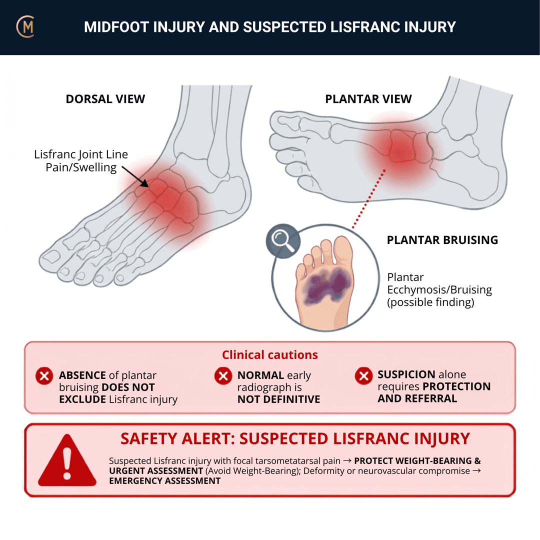
Who: A twist or an axial load — a landing, a step-off, a foot that got trodden on, a fall onto a plantarflexed foot.
What: Ligamentous injury of the tarsometatarsal complex, with or without fracture, with or without instability.
How it behaves: Midfoot pain and swelling. Difficulty weight-bearing or pushing off. Worse on stairs, pivoting and uneven ground. Often described as "my foot felt like it folded."
The one thing to take from this page:
"Plantar bruise = Lisfranc until proven otherwise." And a normal plain film is not a normal midfoot.
Why this one gets missed
| The problem | The consequence |
|---|---|
| Low-velocity injuries are commonly subtle | The story sounds benign |
| Plain films may miss subtle injury | A normal report reassures the wrong way |
| Radiographic criteria vary — no single universally applied rule [1] | There is no clean number to hide behind |
| Delayed diagnosis | Chronic pain · arch collapse · post-traumatic arthritis · poor push-off |
⚠️ No guideline exists. And the systematic review of radiographic criteria finds no single universally applied rule — that is the finding, not a gap in the research. Clinical suspicion carries this decision.
2 · SE — THE SUBJECTIVE PATTERN (the 70%)
Acute trauma, so your course reorders it: MAIN PROBLEM → BODY CHART → HISTORY → BEHAVIOUR.
MAIN PROBLEM
"I landed awkwardly and my foot felt like it folded."
"I can't push off it."
"The top of my foot swelled up and it's still sore two weeks later."
That last sentence — a "sprain" that isn't settling at two weeks — is a re-imaging conversation.
BODY CHART
- Mark the tarsometatarsal joint line specifically, not "the midfoot".
- Draw the plantar arch and look for bruising. If you have not looked at the sole of the foot, you have not assessed this.
- Mark swelling distribution — dorsal midfoot swelling is the pattern.
HISTORY
- Mechanism, in detail — twisting, axial load, a step-off, a fall onto a plantarflexed foot, a direct crush.
- Could they weight-bear immediately, and can they now?
- "Did the foot feel like it folded or gave way?" Ask it in those words.
- Has this been imaged already, and what did the report actually say? A normal plain film does not close the question.
BEHAVIOUR
- Worse on stairs, pivoting and uneven ground — the loading positions that stress the complex.
- Eased by rest, worse on walking.
- Severity: VAS 0–10 at best / at worst / right now.
- Reduced push-off power as a functional complaint.
SPECIAL / SCREENING QUESTIONS
- Any sense of instability or the arch dropping since?
- Is it getting better, plateaued, or worse? Plateau after a "sprain" in this region is a red flag, not a slow responder.
Assessing this region? Download the Ankle & Foot assessment template →
3 · HYPOTHESIS RANKING
| Competing pattern | The one discriminating feature |
|---|---|
| Stress fracture (navicular or metatarsal base) | Gradual load-related pain persisting after activity, localised bony tenderness, positive hop or percussion. Lisfranc is a twist or axial event. |
| Tibialis posterior tendinopathy | Gradual medial arch ache with navicular drop and a failed single-leg heel raise. |
| Cuboid syndrome | Lateral midfoot ache after an inversion sprain, tender cuboid, improves after manipulation. |
And the reason the ranking runs differently here
On most patterns the ranking tells you what to treat. Here it tells you what to exclude before you treat anything. A midfoot injury with a plausible mechanism and focal tarsometatarsal findings is a stability question first and a diagnosis second — because the cost of getting it wrong is not a slower recovery, it is arch collapse and post-traumatic arthritis.
4 · RED FLAGS — DON'T MISS THIS
This section is free, on every pattern, forever. Safety is never gated.
The escalation set
| Finding | What it means | What you do |
|---|---|---|
| Plantar arch bruising (Lisfranc sign) | Ligamentous disruption until proven otherwise | Protect and image |
| Focal tarsometatarsal tenderness after a twist or axial load | The core pattern | Protect and image |
| Pain on forefoot abduction / pronation stress or torsion | Instability | Protect and image |
| Inability to weight-bear | Higher-grade injury | Protect and image |
| Reduced propulsion or push-off power | Functional instability | Protect and image |
| A "sprain" not settling, or plateaued | Missed injury | Re-image — CT or MRI |
| Visible deformity, open injury or neurovascular compromise | Fracture-dislocation | 🚑 Emergency department, now |
| Midfoot widening without deformity | Displacement | Urgent orthopaedic review |
The mismatch rule
When the story sounds benign and the findings don't, immobilise and re-image. Low-velocity injuries are commonly subtle, and the gap between a mild history and focal tarsometatarsal findings is exactly the situation this injury gets missed in.
And two rules that survive everything else
1. A normal plain film is not a normal midfoot. CT and MRI improve detection when clinical suspicion remains high despite equivocal radiographs [2].
2. Protected weight-bearing is the default while the question is open. You are not delaying rehabilitation; you are preventing displacement.
Cross-links: Stress fracture — metatarsal, navicular, calcaneal · Lateral ankle sprain · Tibialis posterior tendinopathy
5 · OE — THE OBJECTIVE (the 30%)
Members
Sections 5 to 7 are for members
The objective exam, management and pearls, plus the six-minute lesson and the debrief. Free with an account.
Sign in or join free6 · MX — MANAGEMENT ESSENTIALS
Members
Sections 5 to 7 are for members
The objective exam, management and pearls, plus the six-minute lesson and the debrief. Free with an account.
Sign in or join free7 · CLINICAL PEARLS + RETRIEVAL CHECK
Members
Sections 5 to 7 are for members
The objective exam, management and pearls, plus the six-minute lesson and the debrief. Free with an account.
Sign in or join freeMembers
The 5-minute lesson is for members
Twelve slides on this pattern: what you'll hear, what to examine, what to do. Free with an account.
Sign in or join freeGuideline anchor
No guideline exists for Lisfranc injury, in Australia or anywhere.
More
Anchored to the current diagnostic and management review (Hammad et al. 2026, PMID 41522288), the systematic review of radiographic diagnostic criteria (Seow et al. 2023, PMID 38012651), and the randomised trial of operative versus non-operative care in non-displaced injuries (Ponkilainen et al. 2025, PMID 39584430).
There is no guideline and — more importantly — the systematic review of radiographic criteria finds no single universally applied rule for diagnosing this injury. That is the finding, not a gap in this page's research. Clinical suspicion and imaging escalation carry the decision, and the page says so.
Members
The evidence summary is for members
Verified diagnostic accuracy, where the evidence stands and what it doesn't settle. Free with an account.
Sign in or join freeSources · 18
ShowHide
Every source on this page resolves and title-verifies against PubMed. Newest first.
- 2023Seow et al. (2023). Inconsistent radiographic diagnostic criteria for lisfranc injuries: a systematic review. BMC musculoskeletal disorders, 24(1), 915. https://doi.org/10.1186/s12891-023-07043-z
- 2024Tang et al. (2024). Comparison of diagnostic performance of X‑ray, CT and MRI in patients with surgically confirmed subtle Lisfranc injuries. Experimental and therapeutic medicine, 27(4), 174. https://doi.org/10.3892/etm.2024.12462
- 2009Raikin et al. (2009). Prediction of midfoot instability in the subtle Lisfranc injury. Comparison of magnetic resonance imaging with intraoperative findings. The Journal of bone and joint surgery. American volume, 91(4), 892-9. https://doi.org/10.2106/JBJS.H.01075
- 2021Ponkilainen et al. (2021). Outcomes after nonoperatively treated non-displaced Lisfranc injury: a retrospective case series of 55 patients. Archives of orthopaedic and trauma surgery, 141(8), 1311-1317. https://doi.org/10.1007/s00402-020-03599-w
- 2025Ponkilainen et al. (2025). Operative versus non-operative treatment for non-displaced Lisfranc injuries: A two-center randomized clinical trial. Scandinavian journal of surgery : SJS : official organ for the Finnish Surgical Society and the Scandinavian Surgical Society, 114(2), 153-161. https://doi.org/10.1177/14574969241295585
- 2002Nunley & Vertullo (2002). Classification, investigation, and management of midfoot sprains: Lisfranc injuries in the athlete. The American journal of sports medicine, 30(6), 871-8. https://doi.org/10.1177/03635465020300061901
- 2006Ly & Coetzee (2006). Treatment of primarily ligamentous Lisfranc joint injuries: primary arthrodesis compared with open reduction and internal fixation. A prospective, randomized study. The Journal of bone and joint surgery. American volume, 88(3), 514-20. https://doi.org/10.2106/JBJS.E.00228
- 2010Stavlas et al. (2010). The role of reduction and internal fixation of Lisfranc fracture-dislocations: a systematic review of the literature. International orthopaedics, 34(8), 1083-91. https://doi.org/10.1007/s00264-010-1101-x
- 2019Han et al. (2019). Comparison of primary arthrodesis versus open reduction with internal fixation for Lisfranc injuries: Systematic review and meta-analysis. Journal of postgraduate medicine, 65(2), 93-100. https://doi.org/10.4103/jpgm.JPGM_414_18
- 2021Attia et al. (2021). Return to Play After Low-Energy Lisfranc Injuries in High-Demand Individuals: A Systematic Review and Meta-Analysis of Athletes and Active Military Personnel. Orthopaedic journal of sports medicine, 9(3), 2325967120988158. https://doi.org/10.1177/2325967120988158
- 2023Talaski et al. (2023). Weightbearing Computed Tomography vs Conventional Tomography for Examination of Varying Degrees of Lisfranc Injures: A Systematic Review of the Literature. Foot & ankle orthopaedics, 8(4), 24730114231209767. https://doi.org/10.1177/24730114231209767
- 2024O'Connor et al. (2024). Primary arthrodesis versus open reduction internal fixation for acute Lisfranc injuries: a systematic review and meta-analysis. Archives of orthopaedic and trauma surgery, 145(1), 49. https://doi.org/10.1007/s00402-024-05700-z
- 2025Rajivan et al. (2025). High rates of return to sport following suture button fixation for ligamentous Lisfranc injuries: A systematic review. World journal of orthopedics, 16(3), 97830. https://doi.org/10.5312/wjo.v16.i3.97830
- 2025Ukita et al. (2025). Diagnosis and assessment of subtle Lisfranc injuries: Comparison of computed tomography and weight-bearing radiography. Journal of orthopaedic science : official journal of the Japanese Orthopaedic Association, 30(6), 1133-1136. https://doi.org/10.1016/j.jos.2025.04.004
- 2025Poulsen et al. (2025). Prospective Validation of a Weightbearing CT Stability Threshold for Subtle Lisfranc Injuries. Foot & ankle international, 46(12), 1386-1394. https://doi.org/10.1177/10711007251374674
- 2025Mohamed et al. (2025). From Missed Diagnosis to Optimal Outcomes: A Comprehensive Review of Lisfranc Injuries. Cureus, 17(10), e94615. https://doi.org/10.7759/cureus.94615
- 2026Semelsberger et al. (2026). Diagnostic evaluation and nonoperative management of Lisfranc injuries in athletes. Knee surgery, sports traumatology, arthroscopy : official journal of the ESSKA, 34(9), 3079-3088. https://doi.org/10.1002/ksa.70244
- 2026Hammad et al. (2026). Lisfranc Injuries: Latest Updates on Diagnostics and Management. Translational sports medicine, 2026, 3933956. https://doi.org/10.1155/tsm2/3933956
Mapped to this pattern
Generic measures that also apply
- Borg RPE
Borg Rating of Perceived Exertion Scale (6-20)
perceived exertion · 1 items · 1 min
- GROC
Global Rating of Change Scale
perceived change since baseline · 1 items · 1 min
- NPRS
Numeric Pain Rating Scale
pain intensity · 1 items · 1 min
- PGIC
Patient Global Impression of Change
perceived change since baseline · 1 items · 1 min
- PSFS
Patient-Specific Functional Scale
patient-generated function · 3 items · 2 min
- SANE
Single Assessment Numeric Evaluation
patient-rated function as a percentage of normal · 1 items · 1 min
- VAS
Visual Analogue Scale
pain intensity · 1 items · 1 min
- Borg CR10
Borg Category Ratio 10 Scale
perceived exertion or symptom intensity, ratio scale · 1 items · 1 min
- GAS
Goal Attainment Scaling
individualised goal achievement · 10 min
Did this page do its job?
CliniStudy is in beta. One tap is enough — it decides what gets fixed first.