
1 · SNAPSHOT
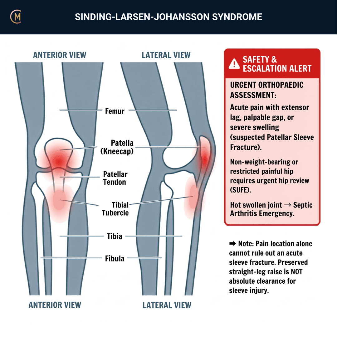
Who: Adolescents in growth spurts. Jumping, sprinting and kicking sports.
What: Traction apophysitis at the inferior pole of the patella.
How it behaves: Gradual anterior knee pain. Worse with stairs, squats, jumping. Point tenderness on the inferior pole.
The one thing to take from this page:
Same site, two very different things. Gradual with full active extension is apophysitis. Acute, with an extensor lag or an inability to straight-leg raise, is a sleeve fracture until imaging says otherwise.
Same site, two stories
| Fits SLJ | Fits patellar sleeve fracture |
|---|---|
| Gradual onset over weeks | Acute, during a jump or kick |
| Load-related, settles with rest | Immediate functional loss |
| Full active extension | Extensor lag, or can't straight-leg raise |
| Local tenderness, no effusion | Major swelling, can't weight bear |
| Deload and reload | Image. Orthopaedic review |
⚠️ There is no clinical guideline for this condition, only a hospital pre-referral note that borrows Osgood-Schlatter management, and no trial evidence for treating it. The management below is imported from Osgood-Schlatter, where the cohort evidence actually sits. That is stated, not hidden.
2 · SE — THE SUBJECTIVE PATTERN (the 70%)
Five stages, in your course's order. The whole subjective is built around one question: is this gradual or is this acute?
MAIN PROBLEM
"It hurts right on the point of my kneecap when I jump."
"Stairs and squats set it off."
"It came on over a few weeks."
BODY CHART
- One finger, on the inferior pole of the patella.
- Mark the tibial tuberosity separately. That is Osgood-Schlatter, and both can coexist.
- Diffuse peri-patellar? Patellofemoral pain.
- Mid-tendon in a skeletally mature athlete? Patellar tendinopathy.
BEHAVIOUR
| Fits SLJ | Points elsewhere | |
|---|---|---|
| Aggs | Jumping, kicking, sprinting, stairs, squats | Prolonged sitting, diffuse → PFP |
| Severity | VAS 0–10 at best / at worst / right now | — |
| Onset | Gradual, over weeks, tracking load and growth | Acute, during a single jump → sleeve fracture |
| Extension | Full active extension | Extensor lag, can't SLR → sleeve fracture |
| Swelling | Local, minor | Major swelling, can't weight bear → escalate |
| Night | Sore after activity | Night or rest pain → screen |
HISTORY
- Gradual or sudden? Ask it directly and do not accept "a few weeks" without checking whether something happened in between.
- Growth spurt? Ask the parent.
- Sport, volume, recent change, number of teams.
- Any moment where it changed suddenly? A sleeve avulsion has been documented occurring in an adolescent who already had SLJ [1]. A known diagnosis does not protect them from the new one.
SPECIAL / SCREENING QUESTIONS
- Can they straight-leg raise? Ask before you examine, then check it.
- Night pain, rest pain, fever, weight loss? Adolescent, bone pain — ask every time.
- Any locking, giving way, or true effusion?
- Hip or groin pain, or a limp? SCFE and Perthes present as knee pain.
Assessing this region? Download the Knee assessment template →
3 · HYPOTHESIS RANKING
| Competing pattern | The one discriminating feature |
|---|---|
| Osgood-Schlatter disease | Tenderness and swelling at the tibial tuberosity rather than the inferior pole. Often coexists. |
| Patellar tendinopathy | Skeletally mature. Same site, tendon rather than apophysis, positive decline squat. |
| Patellofemoral pain | Diffuse rather than point tenderness, poor hip-knee control on step-down. |
| Bipartite patella and other non-traumatic patellar appearances | An imaging finding that mimics pathology — the paediatric patella has several [2]. |
| Referred hip pathology | Screen the hip in every adolescent knee. SCFE and Perthes present here. |
The one that outranks everything on this page
Patellar sleeve fracture. Same site, same age group, completely different pathway. A comparative radiographic study exists precisely because separating paediatric patellar sleeve fractures from SLJ and other inferior-pole pathologies is a recognised clinical problem [3], and the case literature runs in both directions: acute SLJ mimicking a sleeve fracture [4], and a sleeve avulsion occurring in a patient with SLJ (Schmidt-Hebbel et al. 2020), including a minimally displaced one [5].
Minimally displaced is the trap. It is the version that looks like an unusually bad flare-up.
And the rest of the differential in an adolescent
Night pain, rest pain or systemic symptoms move you off this page entirely [6].
4 · RED FLAGS — DON'T MISS THIS
This section is free, on every pattern, forever. Safety is never gated.
| Finding | What it suggests | What you do |
|---|---|---|
| Extensor lag, or can't straight-leg raise | Patellar sleeve fracture | Image. Orthopaedic review |
| Acute onset during a jump or kick | Sleeve fracture, avulsion | Image |
| Major swelling or a true effusion | Fracture, intra-articular injury | Image |
| Inability to weight bear | Fracture | Image |
| A palpable gap or high-riding patella | Extensor mechanism disruption | Urgent orthopaedic review |
| Night pain or pain at rest | Bone tumour, infection | Image. Medical review |
| Fever, unwell, weight loss | Infection, systemic disease | Medical review, same day |
| Limp with hip or groin pain | SCFE, Perthes | Image the hip. Urgent |
The rule: gradual inferior-pole pain with full active extension and a clean systemic screen is yours. An extensor lag, an acute onset, or an inability to straight-leg raise is a sleeve fracture until imaging says otherwise.
Cross-links: Osgood-Schlatter disease · Patellofemoral pain · Sever's disease
5 · OE — THE OBJECTIVE (the 30%)
Members
Sections 5 to 7 are for members
The objective exam, management and pearls, plus the six-minute lesson and the debrief. Free with an account.
Sign in or join free6 · MX — MANAGEMENT ESSENTIALS
Members
Sections 5 to 7 are for members
The objective exam, management and pearls, plus the six-minute lesson and the debrief. Free with an account.
Sign in or join free7 · CLINICAL PEARLS + RETRIEVAL CHECK
Members
Sections 5 to 7 are for members
The objective exam, management and pearls, plus the six-minute lesson and the debrief. Free with an account.
Sign in or join freeMembers
The 5-minute lesson is for members
Twelve slides on this pattern: what you'll hear, what to examine, what to do. Free with an account.
Sign in or join freeGuideline anchor
No Australian guideline covers Sinding-Larsen-Johansson syndrome, and no clinical guideline of any nationality does: a Perth Children's Hospital pre-referral note names it, but only borrows Osgood-Schlatter management.
More
Anchored to the scoping review of the condition (Wilczyński et al. 2024, PMID 39346060) for what is and isn't known, to the comparative radiographic study separating paediatric patellar sleeve fractures from inferior-pole pathologies (Devana et al. 2022, PMID 35142232) for the safety branch, and to the paediatric knee-pain review (Kuwabara et al. 2021, PMID 33818701) for the differential.
There is no clinical guideline here. The evidence base is a scoping review, imaging reviews, a comparative radiographic study and a large number of case reports — the scoping review itself is the acknowledgement that the literature is thin. Management on this page is imported from the better-studied sibling pattern, Osgood-Schlatter, and that import is stated rather than hidden.
Members
The evidence summary is for members
Verified diagnostic accuracy, where the evidence stands and what it doesn't settle. Free with an account.
Sign in or join freeSources · 17
ShowHide
Every source on this page resolves and title-verifies against PubMed. Newest first.
- 2020Schmidt-Hebbel et al. (2020). Patellar sleeve avulsion fracture in a patient with Sinding-Larsen-Johansson syndrome: a case report. BMC musculoskeletal disorders, 21(1), 267. https://doi.org/10.1186/s12891-020-03297-z
- 2024Masroori et al. (2024). Patellar Non-Traumatic Pathologies: A Pictorial Review of Radiologic Findings. Diagnostics (Basel, Switzerland), 14(24). https://doi.org/10.3390/diagnostics14242828
- 2022Devana et al. (2022). Clinical and Radiographic Differentiation of Pediatric Patellar Sleeve Fractures and Other Inferior Pole Pathologies. The American journal of sports medicine, 50(4), 977-983. https://doi.org/10.1177/03635465221073995
- 2018Alassaf (2018). Acute presentation of Sinding-Larsen-Johansson disease simulating patella sleeve fracture: A case report. SAGE open medical case reports, 6, 2050313X18799242. https://doi.org/10.1177/2050313X18799242
- 2023Negrão et al. (2023). Sinding-Larsen-Johansson syndrome with minimally displaced patellar sleeve avulsion fracture: a case report with multimodality approach and literature review. Skeletal radiology, 52(7), 1403-1407. https://doi.org/10.1007/s00256-022-04237-9
- 2021Kuwabara et al. (2021). Narrative Review - Knee Pain in the Pediatric Athlete. Current reviews in musculoskeletal medicine, 14(3), 239-245. https://doi.org/10.1007/s12178-021-09708-5
- 2009Iwamoto et al. (2009). Radiographic abnormalities of the inferior pole of the patella in juvenile athletes. The Keio journal of medicine, 58(1), 50-3. https://doi.org/10.2302/kjm.58.50
- 1989De Flaviis et al. (1989). Ultrasonic diagnosis of Osgood-Schlatter and Sinding-Larsen-Johansson diseases of the knee. Skeletal radiology, 18(3), 193-7. https://doi.org/10.1007/BF00360969
- 2008Draghi et al. (2008). Overload syndromes of the knee in adolescents: Sonographic findings. Journal of ultrasound, 11(4), 151-7. https://doi.org/10.1016/j.jus.2008.09.001
- 2012Valentino et al. (2012). Sinding-Larsen-Johansson syndrome: A case report. Journal of ultrasound, 15(2), 127-9. https://doi.org/10.1016/j.jus.2012.03.001
- 2024Yaya-Quezada et al. (2024). Imaging of the Pediatric Knee. Seminars in musculoskeletal radiology, 28(4), 462-476. https://doi.org/10.1055/s-0044-1786152
- 2010Nath & Lattin GE (2010). Patellar sleeve fracture. Pediatric radiology, 40 Suppl 1, S53. https://doi.org/10.1007/s00247-010-1721-8
- 2014Sullivan et al. (2014). Patellar sleeve fracture. The western journal of emergency medicine, 15(7), 883-4. https://doi.org/10.5811/westjem.2014.8.22265
- 2026Wedderkopp et al. (2026). Incidence of and Risk Factors for Lower Extremity Apophysitis in Children and Adolescents. Sports medicine (Auckland, N.Z.), 56(3), 793-803. https://doi.org/10.1007/s40279-025-02328-w
- 2025Alhamzah et al. (2025). Beyond conservative management: Surgical excision for symptomatic Sinding-Larsen-Johansson syndrome: A case report and literature review. Medicine, 104(52), e46792. https://doi.org/10.1097/MD.0000000000046792
- 2019Malherbe (2019). Traction apophysitis of the knee: A case report. Radiology case reports, 14(1), 18-21. https://doi.org/10.1016/j.radcr.2018.09.012
- 2024Wilczyński et al. (2024). Sinding-Larsen-Johansson disease. Clinical features, imaging findings, conservative treatments and research perspectives: a scoping review. PeerJ, 12, e17996. https://doi.org/10.7717/peerj.17996
Mapped to this pattern
Generic measures that also apply
- Borg RPE
Borg Rating of Perceived Exertion Scale (6-20)
perceived exertion · 1 items · 1 min
- GROC
Global Rating of Change Scale
perceived change since baseline · 1 items · 1 min
- NPRS
Numeric Pain Rating Scale
pain intensity · 1 items · 1 min
- PGIC
Patient Global Impression of Change
perceived change since baseline · 1 items · 1 min
- PSFS
Patient-Specific Functional Scale
patient-generated function · 3 items · 2 min
- SANE
Single Assessment Numeric Evaluation
patient-rated function as a percentage of normal · 1 items · 1 min
- VAS
Visual Analogue Scale
pain intensity · 1 items · 1 min
- Borg CR10
Borg Category Ratio 10 Scale
perceived exertion or symptom intensity, ratio scale · 1 items · 1 min
- GAS
Goal Attainment Scaling
individualised goal achievement · 10 min
Did this page do its job?
CliniStudy is in beta. One tap is enough — it decides what gets fixed first.