
1 · SNAPSHOT
Usually the sequel to something else, and usually missed because everyone stopped looking when the fracture healed.
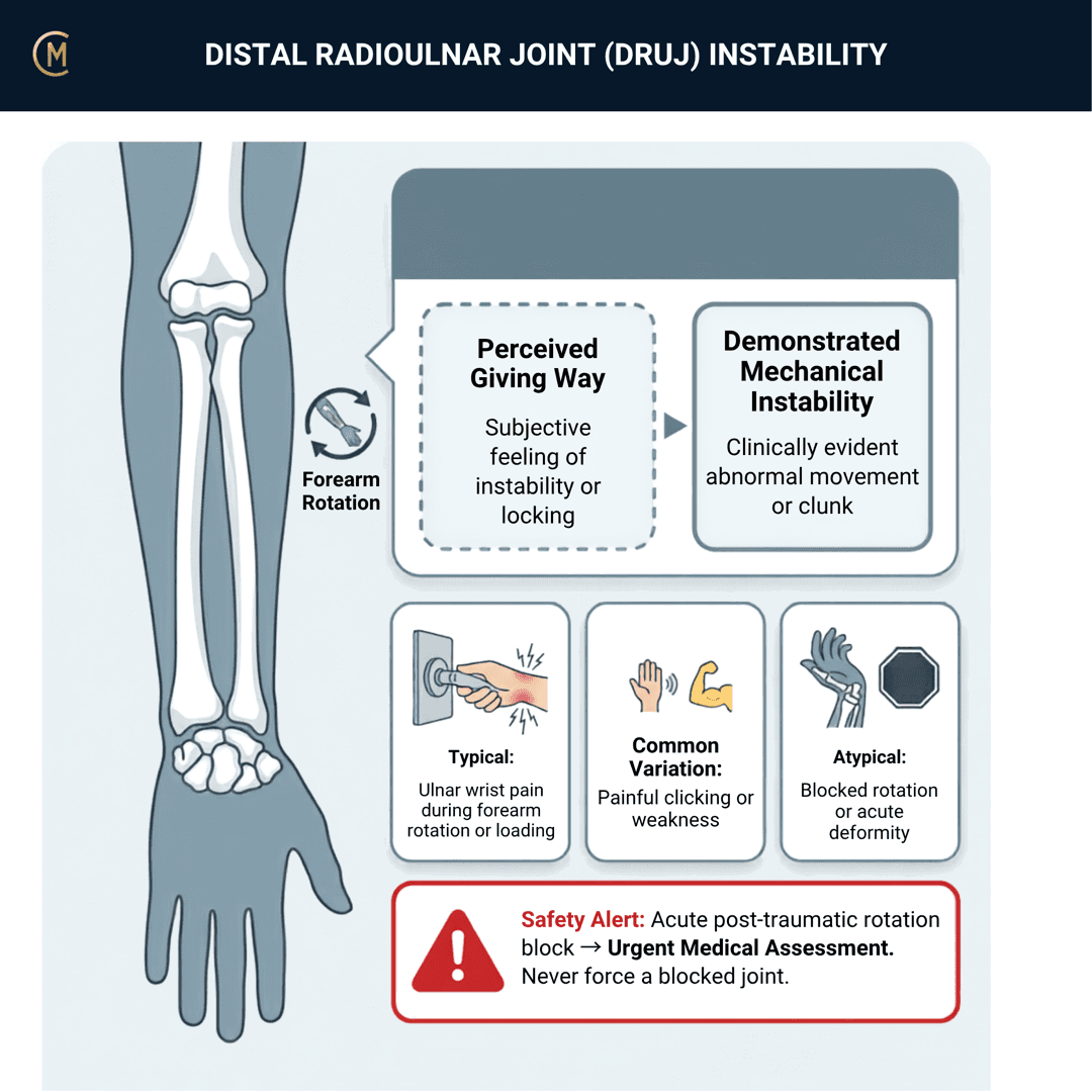
Illness script in one line: ulnar-sided wrist pain with a sense of the forearm giving way when turning a handle, opening a door or lifting a full kettle — often months after a distal radius fracture — with weakness on rotation rather than a clear painful arc.
Caseload frequency: uncommon as a primary referral, common as an unaddressed finding on a wrist you were sent for something else.
The commonest route in is a distal radius fracture that healed, and nobody looked
This is the organising idea: often post-traumatic or post-distal radius fracture.
Read further: DRUJ instability in distal radius fracture [1], predictive factors after surgical treatment of distal radius fracture [2], arthroscopic capsular suture in that setting [3].
Why that matters to a student: the fracture had a diagnosis, a treatment and a discharge. The ulnar-sided pain that follows gets filed as stiffness, and the person best placed to notice it is the physiotherapist doing the wrist rehabilitation — which is you.
| The way it usually goes | What should happen |
|---|---|
| "Wrist fracture, six weeks in plaster, now stiff" | Check DRUJ stability before you write the stiffness programme |
| Rotation is limited, so mobilise rotation | Rotation limited by pain and apprehension is different from rotation limited by stiffness |
| Discharge when range returns | Range can return while the joint stays unstable |
Read this page alongside the distal radius fracture page and the TFCC page's second red flag, for exactly this reason.
2 · SE — THE SUBJECTIVE PATTERN (the 70%)
Main problem
"My wrist gives way when I turn things." medial wrist pain + instability turning handles or lifting.
The word to listen for is gives way, and the movement to listen for is rotation. A patient describing a wrist that hurts is describing something else; a patient describing a forearm they can't trust under rotation is describing this.
Body chart
Ulnar side, over the DRUJ itself — between the ulnar head and the sigmoid notch, dorsally. Ask them to point. Tenderness further distal, at the fovea, is the TFCC page; tenderness over the ECU groove is the ECU page. All three sit on the same ulnar-sided differential.
Behaviour
Typical: weak with pron/sup tasks. Possible click or clunk.
Aggs: turning a door handle or a key, opening a jar, using a screwdriver, pushing up out of a chair, carrying a full kettle or a saucepan, wringing.
Eases: avoiding rotation, a wrist brace, keeping the forearm neutral.
Ask specifically about the quality of the noise. A click is common and often nothing. A clunk with a sense of something moving is different — and the distinction runs the same way as on the TFCC page.
History
Ask the question this page exists for: have you ever broken this wrist?
Then: how was it treated, how long was it immobilised, and when did the ulnar-sided symptoms start? Symptoms that began as the plaster came off, and never fully settled, are the story.
Also ask about: a fall onto an outstretched or twisting hand; any previous wrist surgery; and whether the other wrist feels the same, because generalised laxity changes how you read your own examination.
Special / screening questions
- Any acute event with obvious deformity or an inability to rotate at all? Section 4.
- Any numbness or tingling? Shouldn't be part of this.
- Occupational and sporting rotation load — trades, hairdressing, racquet, gymnastics, weight-bearing through the hand.
- Hypermobility elsewhere? It changes what "increased translation" means.
Assessing this region? Download the Wrist & Hand assessment template →
3 · HYPOTHESIS RANKING
The ulnar-sided wrist pain differential gives the top three.
| Pattern | The one discriminating feature |
|---|---|
| DRUJ instability | A sense of the forearm giving way on rotation, increased translation on ballottement compared with the other side, weakness rather than a painful arc. |
| TFCC injury | Pain rather than instability, tender at the fovea, provoked by ulnar deviation and axial load. Its own page — and the two genuinely coexist. |
| ECU tendinopathy / subluxation | A palpable snap over the ulnar groove on supination, pain on resisted ECU. Its own page. MRI analysis of the ECU tendon and DRUJ has been done together [4] — they are neighbours in every sense. |
| Ulnocarpal impaction | Gradual, grip- and pronation-dominant, load-related rather than unstable. Covered on the TFCC page. |
| Malunited distal radius fracture | Deformity, a changed ulnar variance, and rotation limited by bone rather than by apprehension. Section 4. |
| Acute DRUJ dislocation | An event, obvious deformity, and an inability to rotate. Section 4. |
The discriminator that does most of the work: is the complaint pain or instability — and is there a fracture in the history.
4 · RED FLAGS — DON'T MISS THIS
Free forever. Never gated.
1. Acute DRUJ dislocation is a reduction, not a rehabilitation
A wrist that cannot rotate after an injury, with an obviously prominent ulnar head or a locked forearm, is a dislocated DRUJ until proven otherwise.
Read further: treatment options for acute isolated DRUJ dislocation [5], an adolescent case with a systematic review [6], ulnar head fracture with volar dislocation [7], conservative versus surgical care in acute volar dislocation [8].
Volar dislocations in particular are easy to miss and lock the forearm in supination.
Action: same-day imaging and an orthopaedic opinion. Do not attempt to restore rotation.
2. Frank instability is a referral before it is a programme
Same rule as the TFCC page, and it is worth repeating because a student will meet it there first.
A clunk rather than a click; an ulnar head that visibly or palpably rides dorsally; obvious asymmetry on the piano-key sign; and a patient who says the forearm gives way rather than hurts. Refer before you progress rotation loading, not after it fails.
3. The fracture that didn't heal straight
Rotation limited by bony block, visible deformity, or a changed ulnar variance is a malunion. Predictive factors for DRUJ instability after surgical treatment of a distal radius fracture have been examined (Li et al. 2023) and negative ulnar variance reported to lessen instability after DRUJ disruption in a biomechanical analysis [9], with sigmoid notch morphology analysed on MRI [10].
What that means: the bone geometry matters, you cannot rehabilitate it, and a wrist that won't rotate because of shape needs a surgical opinion rather than more mobilisation.
4. Post-surgical problems belong to the surgeon
A patient deteriorating after DRUJ surgery goes back, not into a programme. Read further: complications of DRUJ implants [11].
5 · OE — THE OBJECTIVE (the 30%)
Members
Sections 5 to 7 are for members
The objective exam, management and pearls, plus the six-minute lesson and the debrief. Free with an account.
Sign in or join free6 · MX — MANAGEMENT ESSENTIALS
Members
Sections 5 to 7 are for members
The objective exam, management and pearls, plus the six-minute lesson and the debrief. Free with an account.
Sign in or join free7 · CLINICAL PEARLS + RETRIEVAL CHECK
Members
Sections 5 to 7 are for members
The objective exam, management and pearls, plus the six-minute lesson and the debrief. Free with an account.
Sign in or join freeMembers
The 4-minute lesson is for members
Twelve slides on this pattern: what you'll hear, what to examine, what to do. Free with an account.
Sign in or join freeGuideline anchor
No Australian guideline and no clinical practice guideline of any kind.
More
The clinical anchors are three reviews of distal radioulnar joint instability (Maclean et al. 2025, PMID 40842425; Kolovich et al. 2024, PMID 39150287; Marès et al. 2023, PMID 36942792) and a comprehensive review of advancements in diagnosis and management (Dmour et al. 2024, PMID 39338197). The anchor that matters most to a physiotherapist is a study of DRUJ instability in distal radius fracture (Huang et al. 2025, PMID 40988534), with predictive factors after surgical treatment of distal radius fracture reported separately (Li et al. 2023, PMID 38050192).
None of these is a physiotherapy document. They are surgical and radiological reviews. There is no rehabilitation guideline for this condition, and the conservative literature is close to absent.
Members
The evidence summary is for members
Verified diagnostic accuracy, where the evidence stands and what it doesn't settle. Free with an account.
Sign in or join freeSources · 20
ShowHide
Every source on this page resolves and title-verifies against PubMed. Newest first.
- 2025Huang et al. (2025). Distal Radioulnar Joint Instability in Distal Radius Fracture. The journal of hand surgery Asian-Pacific volume, 30(6), 571-581. https://doi.org/10.1142/S2424835525400090
- 2023Li et al. (2023). Predictive factors of distal radioulnar joint instability after surgical treatment of distal radius fractures. Medicine, 102(48), e36505. https://doi.org/10.1097/MD.0000000000036505
- 2025Jiang et al. (2025). Arthroscopic capsular suture benefits the clinical prognosis of distal radius fracture with DRUJ instability. BMC surgery, 25(1), 327. https://doi.org/10.1186/s12893-025-03053-x
- 2018Santo et al. (2018). Magnetic resonance imaging analysis of the extensor carpi ulnaris tendon and distal radioulnar joint in triangular fibrocartilage complex tears. Journal of orthopaedic science : official journal of the Japanese Orthopaedic Association, 23(6), 953-958. https://doi.org/10.1016/j.jos.2018.05.009
- 2024Zampetakis et al. (2024). Systematic Review of Acute Isolated Distal Radioulnar Joint Dislocation: Treatment Options. Journal of clinical medicine, 13(24). https://doi.org/10.3390/jcm13247817
- 2025Murphy et al. (2025). Isolated Distal Radioulnar Joint Dislocation in an Adolescent: A Case Report and Systematic Review. Journal of orthopaedic case reports, 15(5), 20-24. https://doi.org/10.13107/jocr.2025.v15.i05.5540
- 2024Samy et al. (2024). Ulnar Head Fracture with Volar Dislocation of the Distal Radioulnar Joint: A Case Series and Systematic Review. Journal of wrist surgery, 13(6), 584-588. https://doi.org/10.1055/s-0043-1777019
- 2023Köroglu et al. (2023). Acute isolated volar distal radioulnar joint dislocation: first surgery or conservative?. Trauma case reports, 48, 100952. https://doi.org/10.1016/j.tcr.2023.100952
- 2024Huddleston et al. (2024). Negative Ulnar Variance Lessens DRUJ Instability After DRUJ Disruption: A Biomechanical Analysis. Hand (New York, N.Y.), 19(3), 448-455. https://doi.org/10.1177/15589447221124233
- 2023Mauler et al. (2023). Midsectional Magnetic Resonance Imaging Analysis of the Sigmoid Notch of the Distal Radioulnar Joint. Journal of wrist surgery, 12(2), 170-176. https://doi.org/10.1055/s-0042-1750874
- 2025Stougie et al. (2025). Aptis Distal Radioulnar Joint Implant: Management of Remarkable Complications. Journal of wrist surgery, 14(2), 134-143. https://doi.org/10.1055/s-0044-1779448
- 2025Maclean (2025). Distal Radioulnar Joint Instability. The journal of hand surgery Asian-Pacific volume, 30(5), 447-457. https://doi.org/10.1142/S2424835525400089
- 2024Kolovich et al. (2024). Distal Radioulnar Joint Instability. Journal of orthopaedic trauma, 38(9S), S4-S10. https://doi.org/10.1097/BOT.0000000000002859
- 2023Marès & Bosch (2023). Distal radioulnar joint instability: Diagnosis and treatment of acute and chronic lesions. Orthopaedics & traumatology, surgery & research : OTSR, 109(1S), 103465. https://doi.org/10.1016/j.otsr.2022.103465
- 2024Dmour et al. (2024). Advancements in Diagnosis and Management of Distal Radioulnar Joint Instability: A Comprehensive Review Including a New Classification for DRUJ Injuries. Journal of personalized medicine, 14(9). https://doi.org/10.3390/jpm14090943
- 2026Hegazy et al. (2026). Open versus arthroscopic management of acute and chronic distal radioulnar joint instability: a systematic review and meta-analysis. BMC musculoskeletal disorders, 27(1). https://doi.org/10.1186/s12891-026-09939-y
- 2023Thomsen & Björkman (2023). Reconstruction of the Distal Oblique Bundle for DRUJ Instability. Journal of wrist surgery, 12(3), 261-264. https://doi.org/10.1055/s-0041-1740488
- 2026Liu & Lim (2026). TFCC Foveal Reconstruction for Chronic DRUJ Instability Using a Single Limb Tendon Graft Technique: A Minimum 32-month Follow-up Study. Journal of wrist surgery, 15(2), 147-153. https://doi.org/10.1055/a-2640-4354
- 2026Challoumas et al. (2026). Efficacy and safety of prosthetic arthroplasty of the distal radioulnar joint: a systematic review. The Journal of hand surgery, European volume, 51(7), 847-855. https://doi.org/10.1177/17531934261415827
- 2024Yeh et al. (2024). Effect of dorsal capsular imbrication on intraoperative DRUJ instability following arthroscopic TFCC repair surgery. BMC musculoskeletal disorders, 25(1), 543. https://doi.org/10.1186/s12891-024-07663-z
Mapped to this pattern
Generic measures that also apply
- Borg RPE
Borg Rating of Perceived Exertion Scale (6-20)
perceived exertion · 1 items · 1 min
- DASH
Disabilities of the Arm, Shoulder and Hand
upper limb disability · 30 items · 8 min
- GROC
Global Rating of Change Scale
perceived change since baseline · 1 items · 1 min
- NPRS
Numeric Pain Rating Scale
pain intensity · 1 items · 1 min
- PGIC
Patient Global Impression of Change
perceived change since baseline · 1 items · 1 min
- PSFS
Patient-Specific Functional Scale
patient-generated function · 3 items · 2 min
- QuickDASH
QuickDASH
upper limb disability, short form · 11 items · 4 min
- SANE
Single Assessment Numeric Evaluation
patient-rated function as a percentage of normal · 1 items · 1 min
- UEFI
Upper Extremity Functional Index
upper limb function · 20 items · 5 min
- VAS
Visual Analogue Scale
pain intensity · 1 items · 1 min
- Borg CR10
Borg Category Ratio 10 Scale
perceived exertion or symptom intensity, ratio scale · 1 items · 1 min
- GAS
Goal Attainment Scaling
individualised goal achievement · 10 min
Did this page do its job?
CliniStudy is in beta. One tap is enough — it decides what gets fixed first.