
1 · SNAPSHOT
Who: Anyone, any sport, any uneven footpath. Highest rates in indoor and court sports, and — a detail worth knowing — higher in females and in children than adults.
What: Sudden inversion, lateral ankle pain, swelling and bruising. Difficulty weight-bearing.
How it behaves: Acutely traumatic, then mechanical. Settles fast in the first fortnight, then plateaus.
The illness script in one line: a sudden inversion injury with lateral swelling and tenderness, where your first job is to rule out fracture, your second is to check it isn't a high ankle sprain, and your third is to stop treating it as trivial.
The numbers that matter:
| Per 1,000 exposures | |
|---|---|
| Females | 13.6 |
| Males | 6.94 |
| Children | 2.85 |
| Adolescents | 1.94 |
| Adults | 0.72 |
| Indoor/court sports | 7 per 1,000 exposures |
([1], 181 prospective studies. No confidence intervals reported. Lateral ankle sprain was the most common type.)
Ankle sprain is roughly 14% of all sport-related injury, and about 80% of those are ligamentous inversion sprains ([2], narrative review — treat that figure as reported, not pooled).
⚠️ No Australian management guideline exists for this presentation; for the X-ray decision, NSW's emergency care protocol uses the Ottawa Ankle Rules. Anchored to the JOSPT Ankle Stability and Movement Coordination Impairments CPG [3], with the Dutch multidisciplinary guideline (Vuurberg 2018) and the ROAST assessment consensus (Delahunt 2018) alongside. The closest Australian material is primary research, not guidance.
Pattern > Label. ATFL versus CFL is not the decision. The decisions are: fracture or not, syndesmosis or not, and how hard are you going to load this.
2 · SE — THE SUBJECTIVE PATTERN (the 70%)
Traumatic onset, so run the UniSA order for trauma: MAIN PROBLEM → BODY CHART → HISTORY → BEHAVIOUR. The mechanism is the story here, so get it before anything else.
MAIN PROBLEM
"What's the main problem today?" — then the two that follow it everywhere in this pattern: can you walk on it, and what do you need to get back to.
BODY CHART
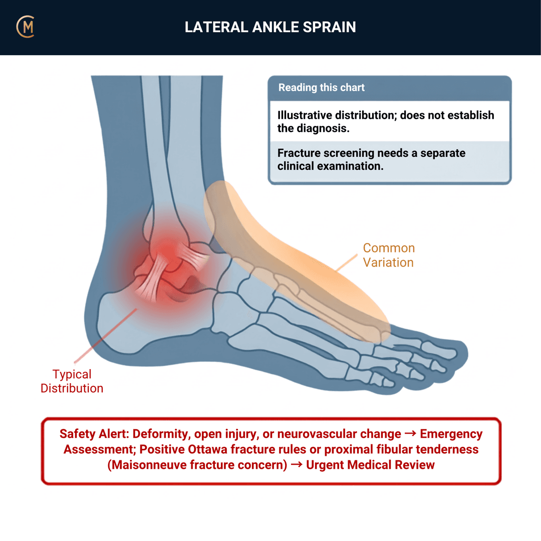
- Lateral ankle, over and anterior to the lateral malleolus — the ATFL. Pain over the CFL sits lower and slightly posterior.
- Map it above the joint line too. Pain proximal to the ankle joint, up the shin, is the syndesmosis question (§3).
- Clear the base of the fifth metatarsal, the navicular, the whole fibula to the head, and the medial malleolus. The proximal fibula is a separate associated-injury assessment, outside the Ottawa ankle/foot rule sites. The chart is a symptom map, not a fracture-screening diagram.
HISTORY — the mechanism does most of the work
- Inversion and plantarflexion → lateral ligaments. This is the common one.
- Dorsiflexion plus external rotation → syndesmosis. Different injury, different timeline, different conversation.
- Ask about the noise, the swelling speed, and whether they kept playing. Immediate marked swelling and an inability to continue is a more severe injury until proven otherwise.
- Previous sprains — this is the single strongest risk factor and the whole reason you're going to bother with rehab [4].
- What footwear, what surface, and had they done anything different that week.
The mechanism worth understanding, because it changes what you tell them — and it is worth saying out loud to the patient, because it reframes blame and builds trust: the injuring motion happens in roughly 50 ms. Peroneal reaction time is 60–90 ms (Fong 2009). The muscle cannot get there in time. So "you should have caught it" and "your ankle is weak" are both wrong — what training changes is what happens before the foot lands, and what happens on the next thousand landings.
BEHAVIOUR
- Aggs: weight-bearing, push-off, uneven ground, inversion, end-range plantarflexion, stairs.
- Eases: rest, elevation, compression, non-weight-bearing.
- Severity and irritability: vigour, severity, time to settle. It sets how much you do today.
SPECIAL / SCREENING QUESTIONS
General health, diabetes, previous ankle surgery, medications, and any neurological symptoms in the foot.
Assessing this region? Download the Ankle & Foot assessment template →
3 · HYPOTHESIS RANKING
Top competing patterns, one discriminating feature each. Rule out the dangerous one first.
| Competing pattern | The one discriminating feature |
|---|---|
| Fracture | Positive applicable Ottawa ankle or foot criteria — apply the separate malleolar/midfoot zones and precise sites in section 4; clinical concern can still warrant imaging |
| Syndesmosis (high ankle) injury | Dorsiflexion–external rotation mechanism, pain above the joint line, and less swelling than the pain would suggest |
| Osteochondral lesion of the talus | Deep ankle pain, catching, and pain that doesn't settle on the expected curve |
| Peroneal tendon injury | Snapping or slipping behind the lateral malleolus, pain on resisted eversion, weak evertors |
| Medial (deltoid) injury | Eversion mechanism or medial tenderness — check for it, don't assume lateral |
The base rates that should change your index of suspicion
It's tempting to treat syndesmosis and osteochondral lesions as edge cases. On 3-T MRI within 10 days of an acute ligamentous ankle injury in 171 injuries [5]:
- Anterior syndesmosis injury in 38%
- Cartilage or osteochondral lesion in 14% (85% of them cartilage rather than osteochondral)
- Lateral ligament injury in 73%
Those are much higher numbers than students expect. A "simple sprain" frequently has something else in it. That doesn't mean scanning everyone — it means the ankle that isn't following the expected recovery curve deserves a second look rather than a longer stretch.
Jones' hypothesis categories that change your plan here: precautions (fracture first, always), sources of symptoms (which structure, and is there more than one), and prognosis (recurrence risk drives your whole plan).
Swelling out of proportion to pain is a lateral sprain. Pain out of proportion to swelling is a syndesmosis until you've checked.
4 · RED FLAGS — DON'T MISS THIS
This section is free, on every pattern, forever.
The Ottawa Ankle Rules — get this exactly right
This is the most useful rule you will learn on placement, and it is a rule-out tool. Nothing more.
Apply the ankle and foot rules separately, within their eligible population:
| Region with pain | Findings supporting radiography assessment |
|---|---|
| Malleolar zone | Bony tenderness at the posterior edge of the distal 6 cm or tip of either malleolus; or inability to take four steps both immediately after injury and at assessment |
| Midfoot zone | Bony tenderness at the navicular or proximal base of the fifth metatarsal; or inability to take four steps both immediately after injury and at assessment |
The fifth-metatarsal base is proximal, not at the distal forefoot. Proximal fibular pain warrants separate assessment for associated injury. Do not force walking to complete the rule. NSW ACI musculoskeletal assessment.
The numbers ([6], 27 studies, 15,581 patients):
| Figure | |
|---|---|
| Pooled −LR, ankle | 0.08 (95% CI 0.03–0.18) |
| Pooled −LR, mid-foot | 0.08 (0.03–0.20) |
| Pooled −LR, children | 0.07 (0.03–0.18) |
| Post-test probability of fracture after a negative rule, at 15% prevalence | under 1.4% |
| Reduction in unnecessary radiographs | 30–40% |
Sensitivity is "almost 100%". Specificity is modest, and the authors deliberately declined to pool it — because the rule was never designed to rule fracture in. A positive rule means image; it does not mean fracture.
And here is the Australian reality check. In an audit of 262 adult ankle X-ray referrals in a South Australian tertiary emergency department, real-world performance was far worse than the pooled figures — overall sensitivity 0.59 (95% CI 0.47–0.71) and specificity 0.37 (0.30–0.44). Physiotherapists had the highest rule compliance at 77.3% and the best accuracy, sensitivity 0.86. Medical officers, registrars and interns were 2.5× more likely to refer for an X-ray despite not meeting the criteria [7].
Read that as encouragement. Applied properly, this rule is one of the places physiotherapy demonstrably outperforms.
Everything else to screen
| Signs / symptoms | Implication |
|---|---|
| Bony tenderness at the proximal fibula | Maisonneuve fracture — palpate the whole fibula, every time; tender means urgent medical review |
| Pain above the joint line, DF–ER mechanism | Syndesmosis injury — §5 |
| Deformity, gross instability, neurovascular compromise | Fracture-dislocation — emergency department, now |
| Hot, red, swollen, systemically unwell (later) | Infection · DVT |
| Numbness, pins and needles, cold foot | Neurovascular injury — assess and document; compromised circulation goes to the emergency department |
Palpate the whole fibula. Every time.
An applicable negative rule lowers the likelihood of the fractures it targets; persistent clinical concern can still require imaging. A positive rule supports imaging assessment, not a fracture diagnosis.
Pain out of proportion to swelling = check the syndesmosis before you rehab the ATFL.
5 · OE — THE OBJECTIVE
Members
Sections 5 to 7 are for members
The objective exam, management and pearls, plus the six-minute lesson and the debrief. Free with an account.
Sign in or join free6 · MX — MANAGEMENT ESSENTIALS
Members
Sections 5 to 7 are for members
The objective exam, management and pearls, plus the six-minute lesson and the debrief. Free with an account.
Sign in or join free7 · CLINICAL PEARLS + RETRIEVAL CHECK
Members
Sections 5 to 7 are for members
The objective exam, management and pearls, plus the six-minute lesson and the debrief. Free with an account.
Sign in or join freeMembers
The 5-minute lesson is for members
Twelve slides on this pattern: what you'll hear, what to examine, what to do. Free with an account.
Sign in or join freeGuideline anchor
No Australian management guideline. For the X-ray decision, NSW's emergency care protocol (Agency for Clinical Innovation) uses the Ottawa Ankle Rules.
More
Management is anchored to the JOSPT Lateral Ankle Ligament Sprains Clinical Practice Guideline, Revision 2021 (Martin et al., PMID 33789434).
No Australian clinical guideline or clinical care standard exists for ankle sprain. Five distinct search strategies returned none. The closest Australian assets are primary studies, not guidelines: Gomes 2020 (South Australian ED audit), Sman 2015 (syndesmosis accuracy, Sydney).
Members
The evidence summary is for members
Verified diagnostic accuracy, where the evidence stands and what it doesn't settle. Free with an account.
Sign in or join freeSources · 22
ShowHide
Every source on this page resolves and title-verifies against PubMed. Newest first.
- 2014Doherty et al. (2014). The incidence and prevalence of ankle sprain injury: a systematic review and meta-analysis of prospective epidemiological studies. Sports medicine (Auckland, N.Z.), 44(1), 123-40. https://doi.org/10.1007/s40279-013-0102-5
- 2009Fong et al. (2009). Understanding acute ankle ligamentous sprain injury in sports. Sports medicine, arthroscopy, rehabilitation, therapy & technology : SMARTT, 1, 14. https://doi.org/10.1186/1758-2555-1-14
- 2021Martin et al. (2021). Ankle Stability and Movement Coordination Impairments: Lateral Ankle Ligament Sprains Revision 2021. The Journal of orthopaedic and sports physical therapy, 51(4), CPG1-CPG80. https://doi.org/10.2519/jospt.2021.0302
- 2019Delahunt & Remus (2019). Risk Factors for Lateral Ankle Sprains and Chronic Ankle Instability. Journal of athletic training, 54(6), 611-616. https://doi.org/10.4085/1062-6050-44-18
- 2025Baltes et al. (2025). The Prevalence, Size, and Anatomic Location of Cartilage and Osteochondral Lesions in Athletes With an Acute Ligamentous Ankle Injury. The American journal of sports medicine, 53(9), 2173-2180. https://doi.org/10.1177/03635465251344187
- 2003Bachmann et al. (2003). Accuracy of Ottawa ankle rules to exclude fractures of the ankle and mid-foot: systematic review. BMJ (Clinical research ed.), 326(7386), 417. https://doi.org/10.1136/bmj.326.7386.417
- 2020Gomes et al. (2020). Adequacy of clinical information in X-ray referrals for traumatic ankle injury with reference to the Ottawa Ankle Rules-a retrospective clinical audit. PeerJ, 8, e10152. https://doi.org/10.7717/peerj.10152
- 2019Bleakley et al. (2019). Rehabilitation Exercises Reduce Reinjury Post Ankle Sprain, But the Content and Parameters of an Optimal Exercise Program Have Yet to Be Established: A Systematic Review and Meta-analysis. Archives of physical medicine and rehabilitation, 100(7), 1367-1375. https://doi.org/10.1016/j.apmr.2018.10.005
- 2017Rivera et al. (2017). Proprioceptive Training for the Prevention of Ankle Sprains: An Evidence-Based Review. Journal of athletic training, 52(11), 1065-1067. https://doi.org/10.4085/1062-6050-52.11.16
- 2009Hupperets et al. (2009). Effect of unsupervised home based proprioceptive training on recurrences of ankle sprain: randomised controlled trial. BMJ (Clinical research ed.), 339, b2684. https://doi.org/10.1136/bmj.b2684
- 2018Bellows & Wong (2018). THE EFFECT OF BRACING AND BALANCE TRAINING ON ANKLE SPRAIN INCIDENCE AMONG ATHLETES: A SYSTEMATIC REVIEW WITH META-ANALYSIS. International journal of sports physical therapy, 13(3), 379-388. PubMed 30038824
- 2019Tassignon et al. (2019). Criteria-Based Return to Sport Decision-Making Following Lateral Ankle Sprain Injury: a Systematic Review and Narrative Synthesis. Sports medicine (Auckland, N.Z.), 49(4), 601-619. https://doi.org/10.1007/s40279-019-01071-3
- 2021Smith et al. (2021). Return to sport decisions after an acute lateral ankle sprain injury: introducing the PAASS framework-an international multidisciplinary consensus. British journal of sports medicine, 55(22), 1270-1276. https://doi.org/10.1136/bjsports-2021-104087
- 2009Lamb et al. (2009). Mechanical supports for acute, severe ankle sprain: a pragmatic, multicentre, randomised controlled trial. Lancet (London, England), 373(9663), 575-81. https://doi.org/10.1016/S0140-6736(09)60206-3
- 2008van Rijn et al. (2008). What is the clinical course of acute ankle sprains? A systematic literature review. The American journal of medicine, 121(4), 324-331.e6. https://doi.org/10.1016/j.amjmed.2007.11.018
- 2016Doherty et al. (2016). Recovery From a First-Time Lateral Ankle Sprain and the Predictors of Chronic Ankle Instability: A Prospective Cohort Analysis. The American journal of sports medicine, 44(4), 995-1003. https://doi.org/10.1177/0363546516628870
- 2018Vuurberg et al. (2018). Diagnosis, treatment and prevention of ankle sprains: update of an evidence-based clinical guideline. British journal of sports medicine, 52(15), 956. https://doi.org/10.1136/bjsports-2017-098106
- 2018Delahunt et al. (2018). Clinical assessment of acute lateral ankle sprain injuries (ROAST): 2019 consensus statement and recommendations of the International Ankle Consortium. British journal of sports medicine, 52(20), 1304-1310. https://doi.org/10.1136/bjsports-2017-098885
- 1996van Dijk et al. (1996). Diagnosis of ligament rupture of the ankle joint. Physical examination, arthrography, stress radiography and sonography compared in 160 patients after inversion trauma. Acta orthopaedica Scandinavica, 67(6), 566-70. https://doi.org/10.3109/17453679608997757
- 2022Beynon et al. (2022). Reliability and validity of physical examination tests for the assessment of ankle instability. Chiropractic & manual therapies, 30(1), 58. https://doi.org/10.1186/s12998-022-00470-0
- 2015Sman et al. (2015). Diagnostic accuracy of clinical tests for ankle syndesmosis injury. British journal of sports medicine, 49(5), 323-9. https://doi.org/10.1136/bjsports-2013-092787
- 2021Netterström-Wedin & Bleakley (2021). Diagnostic accuracy of clinical tests assessing ligamentous injury of the ankle syndesmosis: A systematic review with meta-analysis. Physical therapy in sport : official journal of the Association of Chartered Physiotherapists in Sports Medicine, 49, 214-226. https://doi.org/10.1016/j.ptsp.2021.03.005
Mapped to this pattern
- CAIT
Cumberland Ankle Instability Tool
functional ankle instability · 9 items · 3 min
- FAAM
Foot and Ankle Ability Measure
foot and ankle function · 29 items · 7 min
- SEBT
Star Excursion Balance Test
dynamic single-leg postural control · 10 min
- SLS
Single Leg Stance Test
static single-leg balance · 3 min
- FAOS
Foot and Ankle Outcome Score
foot and ankle outcome · 42 items · 10 min
- Ottawa Ankle
Ottawa Ankle Rules
imaging decision rule · 4 items · 1 min
Generic measures that also apply
- Borg RPE
Borg Rating of Perceived Exertion Scale (6-20)
perceived exertion · 1 items · 1 min
- GROC
Global Rating of Change Scale
perceived change since baseline · 1 items · 1 min
- NPRS
Numeric Pain Rating Scale
pain intensity · 1 items · 1 min
- PGIC
Patient Global Impression of Change
perceived change since baseline · 1 items · 1 min
- PSFS
Patient-Specific Functional Scale
patient-generated function · 3 items · 2 min
- SANE
Single Assessment Numeric Evaluation
patient-rated function as a percentage of normal · 1 items · 1 min
- VAS
Visual Analogue Scale
pain intensity · 1 items · 1 min
- Borg CR10
Borg Category Ratio 10 Scale
perceived exertion or symptom intensity, ratio scale · 1 items · 1 min
- GAS
Goal Attainment Scaling
individualised goal achievement · 10 min
Did this page do its job?
CliniStudy is in beta. One tap is enough — it decides what gets fixed first.