
1 · SNAPSHOT
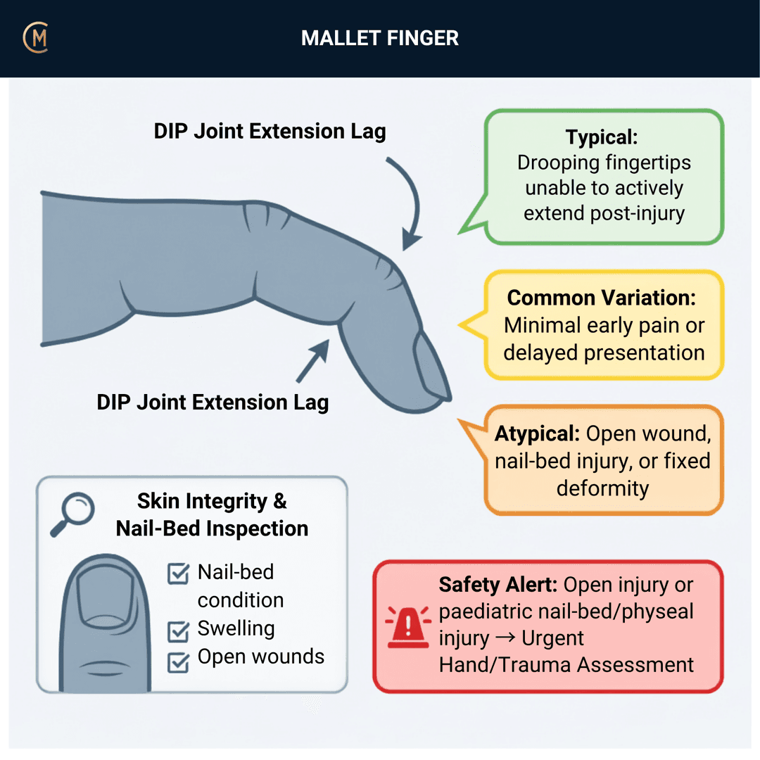
The diagnosis takes five seconds and the treatment takes eight weeks of someone else's discipline.
Illness script in one line: a fingertip took a direct blow — a ball, a crush, a bedsheet, a sock — and the end joint now droops and cannot be actively straightened, though you can straighten it passively without resistance.
Caseload frequency: common, and commonly presenting late. Read further: incidence and management in primary care [1].
The splint is the treatment — and fitting it is your job
This is the whole page. There is no exercise programme, no manual therapy and no progression to design. The intervention is a piece of thermoplastic or plastic on the end of a finger, held in position continuously for six to eight weeks, and the two things that decide the outcome are how well it fits and whether the patient can live with it.
Custom-made finger orthoses have been compared with prefabricated ones and had fewer skin complications, in Archives of Physical Medicine and Rehabilitation [2], and the complications and prognosis of treatment described in the Journal of Hand Surgery [3].
Why that reframes the page: the commonest way splinting fails is not the tendon. It is skin breakdown, maceration, pressure over the dorsum of the DIP, a splint that hurts, and a patient who takes it off. All of those are the responsibility of the person who fitted it. That is you.
| The way students think about it | The way to think about it |
|---|---|
| "Splint for six weeks" — an instruction | A device you fit, check, adjust and re-check |
| Compliance is the patient's problem | Compliance is a fitting problem until proven otherwise |
| Review at six weeks | Review at one week, because that's when the skin tells you |
| The tendon is the treatment target | The skin is the failure point |
2 · SE — THE SUBJECTIVE PATTERN (the 70%)
Main problem
"The end of my finger won't straighten." Sudden pain after fingertip trauma (ball strike, crush). Inability to actively extend DIP joint.
Or, very often, they don't say that at all — May be mistaken for a "jammed finger." Many present a fortnight late, having assumed it would sort itself out. Ask directly: can you straighten the last joint yourself?
Body chart
Dorsal, over the distal phalanx and the DIP joint. Swelling and tenderness at dorsal distal phalanx. Local, not radiating.
Behaviour
Eased by immobilisation. It is not a painful condition after the first few days — which is exactly the problem, because a finger that doesn't hurt is a finger that comes out of the splint.
Aggs: catching the fingertip on things, using the hand normally, gripping.
Eases: the splint.
History
A single event, usually trivial-sounding. The classics are a ball to the fingertip, a crush, and — worth asking about specifically — tucking in a bedsheet or pulling on a sock, because those don't feel like injuries and the patient won't offer them.
How long ago is the question that changes the plan. A late presentation is still worth splinting, but it changes the conversation about what you can expect — see section 6.
Special / screening questions
- Any break in the skin, or damage to the nail or nail fold? Go to section 4 before you go any further.
- How old are they? A mallet posture in a skeletally immature finger is a different problem — section 4. Read further: outcomes of splinting in paediatric mallet finger [4].
- What's their job and their hand dominance? Eight weeks of a splinted index finger is different for a chef, a nurse and a bookkeeper, and the conversation about which splint has to include it.
- Can they physically manage the splint? Read further: patient characteristics affecting outcome [5]. Ask about dexterity, eyesight, whether they live alone, and whether anyone can help.
Assessing this region? Download the Wrist & Hand assessment template →
3 · HYPOTHESIS RANKING
| Pattern | The one discriminating feature |
|---|---|
| Mallet finger (tendinous) | Active DIP extension lost, passive extension full and easy, no bony fragment of consequence on X-ray. |
| Bony mallet (avulsion fracture) | Same clinical picture, a fragment on the film. Doesn't change your day unless the fragment is large or the distal phalanx is subluxed volarly — section 4 [6]. |
| Seymour-type injury in a child | Skeletally immature finger, mallet posture, and a nail-bed or nail-fold injury. That is an open physeal injury until someone says otherwise. Section 4. |
| Chronic mallet with swan-neck | A late presentation where the PIP has gone into hyperextension. Read further: treatment of chronic mallet finger with swan neck deformity [7], reconstruction of chronic soft tissue mallet fingers [8]. Different conversation. |
| DIP joint dislocation or fracture-dislocation | Deformity that isn't passively correctable, or obvious malalignment. If it doesn't straighten passively and easily, it isn't this. |
| Established DIP osteoarthritis | Gradual, no event, bony enlargement, and a stiff joint rather than a drooping one. |
The discriminator that does most of the work: is it passively correctable, and is the skin intact.
4 · RED FLAGS — DON'T MISS THIS
Free forever. Never gated.
Three things belong, and the first one is the one students genuinely miss.
1. A mallet posture in a child with any nail or nail-fold injury
A skeletally immature finger with a mallet posture and a break in the skin, a bleeding nail fold, or a nail that has lifted, is not a simple mallet finger. The growth plate sits under that nail fold, and an injury through it that communicates with the outside is an open fracture — with the infection risk that goes with one.
Read further: outcomes of splinting in paediatric mallet finger [4].
Action: do not splint and review. Refer for same-day medical assessment, and say in the referral that the nail fold is involved. Check this with your educator before the patient leaves.
2. A large bony fragment, or the distal phalanx sitting volar
Most bony mallets are managed exactly like tendinous ones. The exception is a large articular fragment or volar subluxation of the distal phalanx, which is a surgical conversation. Read further: surgical management of delayed mallet finger fractures [6], stack splinting versus Kirschner wire fixation in acute closed Doyle I injuries [9], a single K-wire to prevent poor outcomes in closed soft-tissue injuries [10]. None of it is your decision.
What you do: read the report, and if it mentions a large fragment, joint involvement of a substantial part of the surface, or subluxation, that goes to a hand surgeon before you commit someone to eight weeks.
3. The skin under the splint
This is the red flag that is actually yours to prevent. Read further: complications and prognosis of treatment [3], custom-made versus prefabricated orthoses for skin complications (Witherow et al. 2015).
What to warn them about, in these words: any pain under the splint that wasn't there before, any redness that doesn't fade within a few minutes of a check, any smell, any wetness, or any skin that looks white or dusky — take it off, look, and contact us today. Do not sit on skin pain for a week because you told them the splint has to stay on.
4. Late presentation with a swan-neck deformity
If the PIP has gone into hyperextension, this has moved past a splint-and-review conversation. Read further: treatment of chronic mallet finger with swan neck deformity (Suroto et al. 2023), reconstruction of chronic soft tissue mallet fingers (Demmer et al. 2024). Refer for a hand opinion rather than starting an eight-week trial.
5 · OE — THE OBJECTIVE (the 30%)
Members
Sections 5 to 7 are for members
The objective exam, management and pearls, plus the six-minute lesson and the debrief. Free with an account.
Sign in or join free6 · MX — MANAGEMENT ESSENTIALS
Members
Sections 5 to 7 are for members
The objective exam, management and pearls, plus the six-minute lesson and the debrief. Free with an account.
Sign in or join free7 · CLINICAL PEARLS + RETRIEVAL CHECK
Members
Sections 5 to 7 are for members
The objective exam, management and pearls, plus the six-minute lesson and the debrief. Free with an account.
Sign in or join freeMembers
The 4-minute lesson is for members
Twelve slides on this pattern: what you'll hear, what to examine, what to do. Free with an account.
Sign in or join freeGuideline anchor
No Australian guideline, and no clinical practice guideline of any kind.
More
The clinical anchor is a review of acute mallet finger injuries in the Journal of Hand Surgery (Sivakumar et al. 2023, PMID 36509592) and a current concepts paper in Hand (Alla et al. 2014, PMID 24839413). The treatment-decision anchor is a systematic review and meta-analysis comparing surgical treatment with orthosis splinting (Peng et al. 2023, PMID 36625383). The orthosis anchor — and the one that matters most to a physiotherapist — is a comparison of custom-made and prefabricated finger orthoses for skin complications in Archives of Physical Medicine and Rehabilitation (Witherow et al. 2015, PMID 26163944), with complications and prognosis of treatment described in the Journal of Hand Surgery (Stern et al. 1988, PMID 3379263).
None of these is a clinical practice guideline. The orthosis paper is the closest thing to a physiotherapy-facing evidence anchor for this condition and it is about skin complications, not extension lag.
Members
The evidence summary is for members
Verified diagnostic accuracy, where the evidence stands and what it doesn't settle. Free with an account.
Sign in or join freeSources · 20
ShowHide
Every source on this page resolves and title-verifies against PubMed. Newest first.
- 2024Krastman et al. (2024). Incidence and management of mallet finger in Dutch primary care: a cohort study. BJGP open, 8(1). https://doi.org/10.3399/BJGPO.2023.0040
- 2015Witherow & Peiris (2015). Custom-Made Finger Orthoses Have Fewer Skin Complications Than Prefabricated Finger Orthoses in the Management of Mallet Injury: A Systematic Review and Meta-Analysis. Archives of physical medicine and rehabilitation, 96(10), 1913-1923.e1. https://doi.org/10.1016/j.apmr.2015.04.026
- 1988Stern & Kastrup (1988). Complications and prognosis of treatment of mallet finger. The Journal of hand surgery, 13(3), 329-34. https://doi.org/10.1016/s0363-5023(88)80002-9
- 2018Lin & Samora (2018). Outcomes of Splinting in Pediatric Mallet Finger. The Journal of hand surgery, 43(11), 1041.e1-1041.e9. https://doi.org/10.1016/j.jhsa.2018.03.037
- 2023Azad et al. (2023). A Prospective Analysis of Patient Characteristics Affecting the Outcome of Dorsal Splinting for Soft Tissue Mallet Injuries. Hand (New York, N.Y.), 18(8), 1330-1335. https://doi.org/10.1177/15589447221093674
- 2026Lee et al. (2026). Surgical Management of Delayed Mallet Finger Fractures Using Combined Two-Extension Block Kirschner Wire and Dorsal Counterforce Techniques. The Journal of hand surgery, 51(5), 555-563. https://doi.org/10.1016/j.jhsa.2025.12.019
- 2023Suroto et al. (2023). Treatment of chronic mallet finger with swan neck deformity using a modified Spiral Oblique Retinacular Ligament (SORL) reconstruction procedure: A case series and technical note. International journal of surgery case reports, 104, 107925. https://doi.org/10.1016/j.ijscr.2023.107925
- 2024Demmer et al. (2024). Reconstruction of Chronic Soft Tissue Mallet Fingers: Outcomes of Step-Plasty vs. Purse-String Suture. Journal of functional morphology and kinesiology, 9(3). https://doi.org/10.3390/jfmk9030144
- 2022Velez et al. (2022). Stack Splinting Versus Kirschner Wire Treatment in Acute Closed Mallet Finger Doyle I. Plastic surgery (Oakville, Ont.), 30(2), 117-121. https://doi.org/10.1177/22925503211003838
- 2021Aksan et al. (2021). A single K-wire to prevent poor outcomes in closed soft-tissue mallet finger management due to patient non-compliance. Archives of orthopaedic and trauma surgery, 141(4), 693-698. https://doi.org/10.1007/s00402-021-03793-4
- 2023Sivakumar et al. (2023). Acute Mallet Finger Injuries-A Review. The Journal of hand surgery, 48(3), 283-291. https://doi.org/10.1016/j.jhsa.2022.10.013
- 2014Alla et al. (2014). Current concepts: mallet finger. Hand (New York, N.Y.), 9(2), 138-44. https://doi.org/10.1007/s11552-014-9609-y
- 2026Dinh et al. (2026). Approach to mallet finger injury: Practical guide for Canadian primary care physicians. Canadian family physician Medecin de famille canadien, 72(2), 93-97. https://doi.org/10.46747/cfp.720293
- 2021Ayhan et al. (2021). Evaluating Stack Splint Use for Mallet Finger. The journal of hand surgery Asian-Pacific volume, 26(1), 47-51. https://doi.org/10.1142/S2424835521500077
- 2024Acar (2024). The use of Stack splint or aluminum finger splint in the conservative management of acute Doyle type IVb bony mallet finger. Journal of orthopaedic science : official journal of the Japanese Orthopaedic Association, 29(4), 1091-1096. https://doi.org/10.1016/j.jos.2023.07.020
- 2025Hou et al. (2025). Customized 3-Dimensional-Printed Finger Splints for Mallet Finger. Annals of plastic surgery, 94(3S Suppl 1), S9-S12. https://doi.org/10.1097/SAP.0000000000004190
- 2026Roberts et al. (2026). A simple, rapid technique for preparing and applying a Zimmer finger splint. Annals of the Royal College of Surgeons of England. https://doi.org/10.1308/rcsann.2026.0083
- 2024Kim et al. (2024). Effectiveness of a novel finger range-of-motion brace for extensor tendon injury: a report of 10 patients. BMC musculoskeletal disorders, 25(1), 767. https://doi.org/10.1186/s12891-024-07899-9
- 2023Peng et al. (2023). Comparative outcomes between surgical treatment and orthosis splint for mallet finger: a systematic review and meta-analysis. Journal of plastic surgery and hand surgery, 57(1-6), 54-63. https://doi.org/10.1080/2000656X.2022.2164291
- 2023Slichter et al. (2023). Posttraumatic Osteoarthritis of the Distal Interphalangeal Joint: A Follow-Up Study of 12 Years After Nonsurgical Treatment of Mallet Finger Fractures. The Journal of hand surgery, 48(7), 691-698. https://doi.org/10.1016/j.jhsa.2023.03.027
Generic measures that also apply
- Borg RPE
Borg Rating of Perceived Exertion Scale (6-20)
perceived exertion · 1 items · 1 min
- DASH
Disabilities of the Arm, Shoulder and Hand
upper limb disability · 30 items · 8 min
- GROC
Global Rating of Change Scale
perceived change since baseline · 1 items · 1 min
- NPRS
Numeric Pain Rating Scale
pain intensity · 1 items · 1 min
- PGIC
Patient Global Impression of Change
perceived change since baseline · 1 items · 1 min
- PSFS
Patient-Specific Functional Scale
patient-generated function · 3 items · 2 min
- QuickDASH
QuickDASH
upper limb disability, short form · 11 items · 4 min
- SANE
Single Assessment Numeric Evaluation
patient-rated function as a percentage of normal · 1 items · 1 min
- UEFI
Upper Extremity Functional Index
upper limb function · 20 items · 5 min
- VAS
Visual Analogue Scale
pain intensity · 1 items · 1 min
- Borg CR10
Borg Category Ratio 10 Scale
perceived exertion or symptom intensity, ratio scale · 1 items · 1 min
- GAS
Goal Attainment Scaling
individualised goal achievement · 10 min
Did this page do its job?
CliniStudy is in beta. One tap is enough — it decides what gets fixed first.