
1 · SNAPSHOT
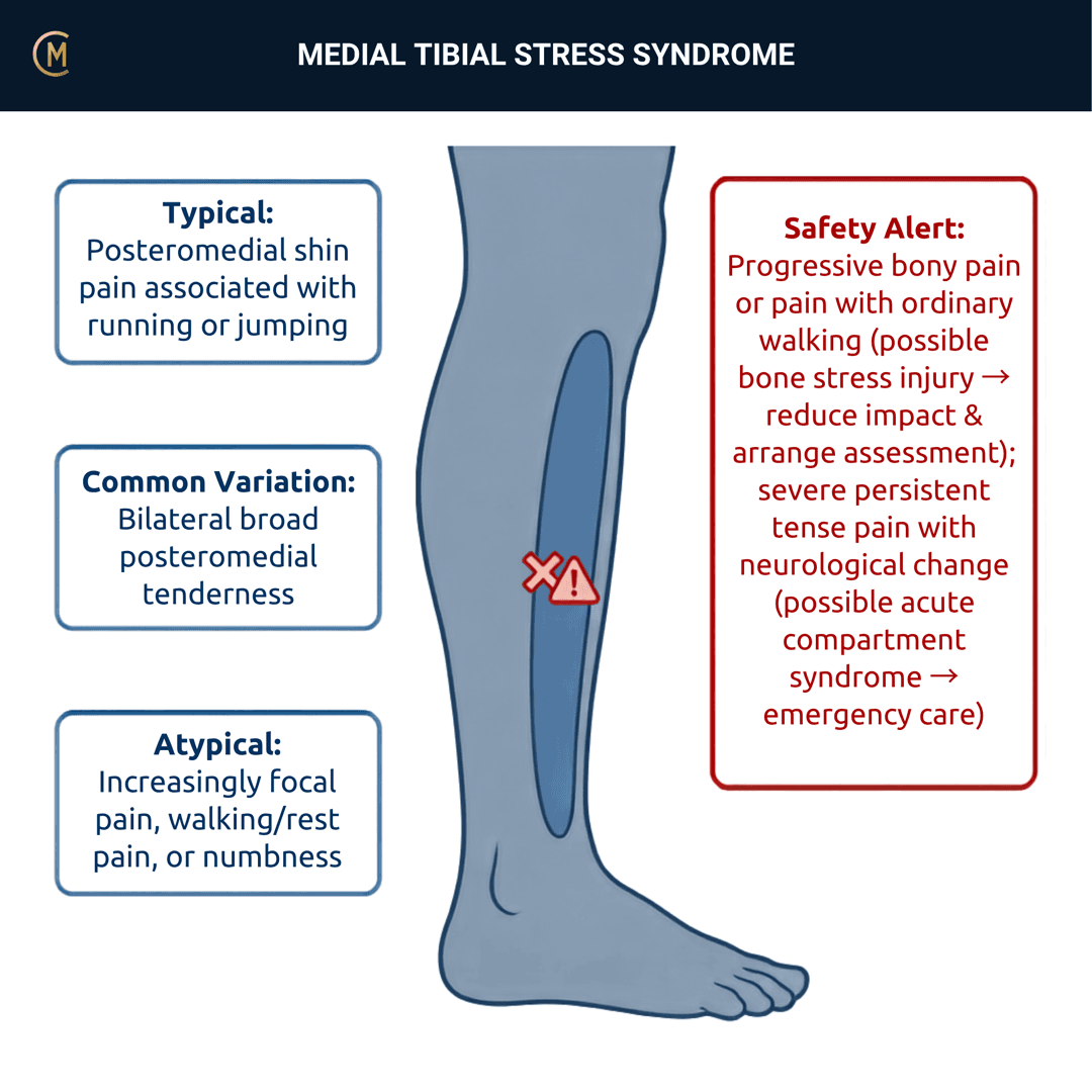
Who: Runners, military recruits, dancers, anyone whose weekly load stepped up. Women more than men, consistently.
What: Diffuse pain along the posteromedial tibial border, brought on by exercise.
How it behaves: Comes on during or after running, settles with rest, comes back at the same distance.
The one thing to take from this page:
There is no clinical practice guideline for this condition. Not in Australia — anywhere.
A PubMed publication-type search across MTSS, shin splints and exertional leg pain returned zero guideline, practice-guideline or consensus records. Everything below is anchored to two systematic reviews, and both of them predate half the trials cited.
How common:
| Population | Incidence |
|---|---|
| 124 UK naval recruits, 10 weeks | 35% — women 53%, men 28%, RR 2.03 |
| 221 PE-teacher students, 10 months (prospective) | 25% — women 39%, men 21% |
| 468 British male infantry recruits, 26 weeks | 7.9% |
| 254 injured novice runners | MTSS was the single most common diagnosis — 15% |
[1][2][3]
And the name is wrong about the mechanism. "Stress syndrome" and "shin splints" read as periostitis from traction. Histological studies fail to provide evidence for that. In 2025 an Australian group proposed dropping the name entirely [4].
Pattern > Label. This one is a label that has actively misled the profession about what the tissue is doing.
2 · SE — THE SUBJECTIVE PATTERN (the 70%)
Five stages, in your course's order. Insidious and load-driven, so BEHAVIOUR before HISTORY — but the load history is where the diagnosis lives.
MAIN PROBLEM
"My shins hurt when I run." Ask immediately: which part, and how big an area?
BODY CHART
- Posteromedial tibial border, distal two-thirds, diffuse over a long stretch.
- Ask them to trace it with one finger. A line they can draw with a fingertip is a different question — §3.
- Anterior shin pain is not this pattern. Say that out loud.
BEHAVIOUR — this is where it separates
- Aggs: running, jumping, hard surfaces, and specifically the same distance each time.
- Eases: stopping. Does it settle within minutes, or does it persist and wake them?
- 24-hr pattern: a distance pattern, not a clock pattern — the same distance each time.
- Night: the page's own question: does it settle within minutes, or persist and wake them? Waking is the bone-stress direction.
- Severity: VAS 0–10 at best / at worst / right now.
- Ask the question that sorts the triad: "Does it hurt when you're not running?"
- Irritability: resting symptoms, vigour, time to settle.
HISTORY
- The load change. New programme, a return after time off, a surface change, a shoe change, a base-training block.
- Have you had this before? Prior MTSS is the strongest thing you can ask about — OR 5.03 (95% CI 1.90–13.30), prospectively, in 221 students [2].
- Smoking, and general aerobic fitness — both were independently associated in 468 prospectively-followed recruits.
SPECIAL / SCREENING QUESTIONS
Energy availability, menstrual history, disordered eating risk, bone health, previous stress fracture, fever, night pain. See §4.
The risk factors — and which ones were actually established prospectively
This distinction is the whole quality of your reasoning here.
| Prospectively established | Estimate |
|---|---|
| Previous MTSS | OR 5.03 (95% CI 1.90–13.30) [2] |
| Female sex | OR 3.14 (1.39–7.11) · and RR 2.03 in recruits |
| Above-average BMI | OR 2.29 (1.02–5.16) |
| Pronated foot type | RR 1.70 |
| Cross-sectional or mixed pooling — association, not risk | Estimate |
|---|---|
| Navicular drop | MD 1.19 mm (95% CI 0.54–1.84) |
| Ankle plantarflexion ROM | MD 5.94° (3.65–8.24) |
| Hip external rotation ROM | MD 3.95° (1.78–6.13) |
| Ankle DORSIflexion ROM | MD −0.01° (−0.96 to 0.93), p = .98 — NOT a risk factor |
| Q-angle | MD −0.22° (−0.95 to 0.50) — not a risk factor |
Read the dorsiflexion row. In the prospective cohort that measured navicular drop and hip range, neither was significant either.
The strongest single question is "have you had this before?" And the second strongest is "what changed?"
Assessing this region? Download the Ankle & Foot assessment template →
3 · HYPOTHESIS RANKING
This differential is the entire clinical value of the page. Four things cause exertional leg pain and they separate on timing and topography, not on a test.
| Pattern | The one discriminating feature |
|---|---|
| MTSS | Diffuse tenderness along the posteromedial border over ≥5 cm, during or after exercise, eases with rest |
| Tibial stress fracture | Focal bone pain, persists at rest and at night, hurts on percussion and with daily walking. Prior stress fracture OR 4.99 (95% CI 2.91–8.56); female sex OR 2.31 (1.24–4.29) |
| ↳ Anterior tibial cortex | Pain over the anterior cortex. The "dreaded black line" — refer, do not manage as MTSS |
| Chronic exertional compartment syndrome | Tightness, ache or paraesthesia at a predictable exercise dose, relieving within minutes of stopping, normal at rest |
| Popliteal artery entrapment | Vascular, not compartmental. Check pulses. The timing does not fit the compartment story |
| DVT | After immobilisation or travel, not exercise-dose dependent |
The ≥5 cm rule — say what it actually is
It is a case definition, not a test.
The criterion — "pain on the posteromedial tibial border during exercise, with pain on palpation of the tibia over a length of at least 5 cm" — originates as a definition [5] and is used as a trial inclusion criterion.
It has no sensitivity, no specificity and no likelihood ratio anywhere in this library. The same is true for the diffuse-versus-focal hop discriminator.
Teach it as the rule the literature uses to define the entity — and say plainly that nobody has tested it. That sentence is worth more to a student than a fabricated number.
What the tissue is actually doing — nobody knows
- Periostitis from traction is not supported by histology. Moen's review states it directly; the proposed mechanism is bone resorption outpacing formation.
- Biopsy evidence: six specimens. Linear microcracks in four (one artefactual, so 50%), no diffuse microdamage, and a near-total absence of a repair reaction [6].
- In 2025 the researchers who built this evidence base proposed renaming it Load Induced Medial-Leg Pain, on the grounds that there is "no conclusive evidence regarding the pathophysiology" and that "tibial stress" gets misread as bone stress injury.
Jones' hypothesis categories that change your plan: pathophysiology — and the honest answer is that it is unresolved — plus precautions (the stress fracture) and contributing factors (load).
A student who can say "nobody knows what this tissue is doing, and here is how I'd sort it from a stress fracture anyway" is reasoning better than one who names a mechanism.
4 · RED FLAGS — DON'T MISS THIS
This section is free, on every pattern, forever.
| Signs / symptoms | Implication |
|---|---|
| Focal bone pain that persists at rest or at night, hurts on percussion and with walking | Tibial stress fracture |
| Pain over the anterior tibial cortex | High-risk stress fracture. Refer — do not manage as MTSS |
| Low energy availability, menstrual disturbance, disordered eating, repeated bone injuries | Relative energy deficiency / bone health. Screen it |
| Escalating pain with tense compartments, neurological symptoms, pain out of proportion | Compartment syndrome — acute is an emergency |
| Calf pain, swelling, warmth after immobilisation or travel | DVT |
| Night pain unchanged by position, systemic features, in a young athlete | Bone tumour |
The high-risk one, by name
The anterior tibial cortex is not a variant of the posteromedial story. It is a different prognosis.
Across the systematic review of high-risk stress fractures of the lower leg [7]: for the anterior tibia there are only 2 studies, N = 31, with "unsatisfying outcomes of conservative therapy", high complication rates and few returning to sport. GRADE: low.
Topography decides this. Anterior cortex pain gets a referral, not a loading programme.
Why imaging will not settle it for you
| Tibial stress reaction on MRI in asymptomatic college distance runners | 43% (21 runners) — and it did not predict future stress reaction or fracture |
| Symptomatic legs in MTSS showing bone marrow or periosteal oedema | only 43.5% (52 athletes) |
| And absence of oedema | associated with a LONGER recovery (p = .033 / .013) |
| Radiograph sensitivity for stress fracture | 12% (95% CI 0–29) to 56% (39–72) |
| MRI sensitivity | 68% (45–90) to 99% (95–100) |
[8][9][10]
A normal X-ray means nothing here. Sensitivity as low as 12%. If you are worried about a stress fracture, an unremarkable plain film is not reassurance.
Focal, at rest, at night, hurts on percussion — that is bone until imaging says otherwise, and a normal X-ray is not imaging saying otherwise.
Anterior tibial cortex pain gets referred, not loaded.
Screen energy availability in anyone with repeated bone injuries.
5 · OE — THE OBJECTIVE (the 30%)
Members
Sections 5 to 7 are for members
The objective exam, management and pearls, plus the six-minute lesson and the debrief. Free with an account.
Sign in or join free6 · MX — MANAGEMENT ESSENTIALS
Members
Sections 5 to 7 are for members
The objective exam, management and pearls, plus the six-minute lesson and the debrief. Free with an account.
Sign in or join free7 · CLINICAL PEARLS + RETRIEVAL CHECK
Members
Sections 5 to 7 are for members
The objective exam, management and pearls, plus the six-minute lesson and the debrief. Free with an account.
Sign in or join freeMembers
The 5-minute lesson is for members
Twelve slides on this pattern: what you'll hear, what to examine, what to do. Free with an account.
Sign in or join freeGuideline anchor
No Australian guideline for this presentation.
More
Anchored to null.
There is no guideline. Both anchors below are systematic reviews, developed without GRADE and without a formal recommendation process, and both predate half the trials cited on this page.
Members
The evidence summary is for members
Verified diagnostic accuracy, where the evidence stands and what it doesn't settle. Free with an account.
Sign in or join freeSources · 27
ShowHide
Every source on this page resolves and title-verifies against PubMed. Newest first.
- 2004Yates & White (2004). The incidence and risk factors in the development of medial tibial stress syndrome among naval recruits. The American journal of sports medicine, 32(3), 772-80. https://doi.org/10.1177/0095399703258776
- 2018Bliekendaal et al. (2018). Incidence and risk factors of medial tibial stress syndrome: a prospective study in Physical Education Teacher Education students. BMJ open sport & exercise medicine, 4(1), e000421. https://doi.org/10.1136/bmjsem-2018-000421
- 2014Nielsen et al. (2014). A prospective study on time to recovery in 254 injured novice runners. PloS one, 9(6), e99877. https://doi.org/10.1371/journal.pone.0099877
- 2025Anderson et al. (2025). Medial Tibial Stress Syndrome Needs a New Name-Make No Bones About It. The Journal of orthopaedic and sports physical therapy, 55(10), 621-622. https://doi.org/10.2519/jospt.2025.13411
- 2009Moen et al. (2009). Medial tibial stress syndrome: a critical review. Sports medicine (Auckland, N.Z.), 39(7), 523-46. https://doi.org/10.2165/00007256-200939070-00002
- 2019Winters et al. (2019). Microcrack-associated bone remodeling is rarely observed in biopsies from athletes with medial tibial stress syndrome. Journal of bone and mineral metabolism, 37(3), 496-502. https://doi.org/10.1007/s00774-018-0945-9
- 2015Mallee et al. (2015). Surgical versus conservative treatment for high-risk stress fractures of the lower leg (anterior tibial cortex, navicular and fifth metatarsal base): a systematic review. British journal of sports medicine, 49(6), 370-6. https://doi.org/10.1136/bjsports-2013-093246
- 2004Bergman et al. (2004). Asymptomatic tibial stress reactions: MRI detection and clinical follow-up in distance runners. AJR. American journal of roentgenology, 183(3), 635-8. https://doi.org/10.2214/ajr.183.3.1830635
- 2014Moen et al. (2014). A prospective study on MRI findings and prognostic factors in athletes with MTSS. Scandinavian journal of medicine & science in sports, 24(1), 204-10. https://doi.org/10.1111/j.1600-0838.2012.01467.x
- 2016Wright et al. (2016). Diagnostic Accuracy of Various Imaging Modalities for Suspected Lower Extremity Stress Fractures: A Systematic Review With Evidence-Based Recommendations for Clinical Practice. The American journal of sports medicine, 44(1), 255-63. https://doi.org/10.1177/0363546515574066
- 2018Winters et al. (2018). Medial tibial stress syndrome can be diagnosed reliably using history and physical examination. British journal of sports medicine, 52(19), 1267-1272. https://doi.org/10.1136/bjsports-2016-097037
- 1995Fredericson et al. (1995). Tibial stress reaction in runners. Correlation of clinical symptoms and scintigraphy with a new magnetic resonance imaging grading system. The American journal of sports medicine, 23(4), 472-81. https://doi.org/10.1177/036354659502300418
- 2012Moen et al. (2012). The treatment of medial tibial stress syndrome in athletes; a randomized clinical trial. Sports medicine, arthroscopy, rehabilitation, therapy & technology : SMARTT, 4, 12. https://doi.org/10.1186/1758-2555-4-12
- 2025Naderi et al. (2025). Effects of Integrating Lower-Leg Exercises Into a Multimodal Therapeutic Approach on Medial Tibial Stress Syndrome Management Among Recreational Runners: A Randomized Controlled Study. Orthopaedic journal of sports medicine, 13(2), 23259671241311849. https://doi.org/10.1177/23259671241311849
- 2013Winters et al. (2013). Treatment of medial tibial stress syndrome: a systematic review. Sports medicine (Auckland, N.Z.), 43(12), 1315-33. https://doi.org/10.1007/s40279-013-0087-0
- 2022Naderi et al. (2022). Foot Orthoses Enhance the Effectiveness of Exercise, Shockwave, and Ice Therapy in the Management of Medial Tibial Stress Syndrome. Clinical journal of sport medicine : official journal of the Canadian Academy of Sport Medicine, 32(3), e251-e260. https://doi.org/10.1097/JSM.0000000000000926
- 2018Bonanno et al. (2018). Effectiveness of foot orthoses for the prevention of lower limb overuse injuries in naval recruits: a randomised controlled trial. British journal of sports medicine, 52(5), 298-302. https://doi.org/10.1136/bjsports-2017-098273
- 2017Gomez Garcia et al. (2017). Shockwave treatment for medial tibial stress syndrome in military cadets: A single-blind randomized controlled trial. International journal of surgery (London, England), 46, 102-109. https://doi.org/10.1016/j.ijsu.2017.08.584
- 2016Barton et al. (2016). Running retraining to treat lower limb injuries: a mixed-methods study of current evidence synthesised with expert opinion. British journal of sports medicine, 50(9), 513-26. https://doi.org/10.1136/bjsports-2015-095278
- 2014Nielsen et al. (2014). Excessive progression in weekly running distance and risk of running-related injuries: an association which varies according to type of injury. The Journal of orthopaedic and sports physical therapy, 44(10), 739-47. https://doi.org/10.2519/jospt.2014.5164
- 2013Newman et al. (2013). Risk factors associated with medial tibial stress syndrome in runners: a systematic review and meta-analysis. Open access journal of sports medicine, 4, 229-41. https://doi.org/10.2147/OAJSM.S39331
- 2015Hamstra-Wright et al. (2015). Risk factors for medial tibial stress syndrome in physically active individuals such as runners and military personnel: a systematic review and meta-analysis. British journal of sports medicine, 49(6), 362-9. https://doi.org/10.1136/bjsports-2014-093462
- 2017Reinking et al. (2017). Medial Tibial Stress Syndrome in Active Individuals: A Systematic Review and Meta-analysis of Risk Factors. Sports health, 9(3), 252-261. https://doi.org/10.1177/1941738116673299
- 2016Winters et al. (2016). The medial tibial stress syndrome score: a new patient-reported outcome measure. British journal of sports medicine, 50(19), 1192-9. https://doi.org/10.1136/bjsports-2015-095060
- 2015Wright et al. (2015). Risk factors associated with lower extremity stress fractures in runners: a systematic review with meta-analysis. British journal of sports medicine, 49(23), 1517-23. https://doi.org/10.1136/bjsports-2015-094828
- 2022Vogels et al. (2022). Clinical Consensus on Diagnosis and Treatment of Patients with Chronic Exertional Compartment Syndrome of the Leg: A Delphi Analysis. Sports medicine (Auckland, N.Z.), 52(12), 3055-3064. https://doi.org/10.1007/s40279-022-01729-5
- 2011Yeung et al. (2011). Interventions for preventing lower limb soft-tissue running injuries. The Cochrane database of systematic reviews, 2011(7), CD001256. https://doi.org/10.1002/14651858.CD001256.pub2
Mapped to this pattern
Generic measures that also apply
- Borg RPE
Borg Rating of Perceived Exertion Scale (6-20)
perceived exertion · 1 items · 1 min
- GROC
Global Rating of Change Scale
perceived change since baseline · 1 items · 1 min
- NPRS
Numeric Pain Rating Scale
pain intensity · 1 items · 1 min
- PGIC
Patient Global Impression of Change
perceived change since baseline · 1 items · 1 min
- PSFS
Patient-Specific Functional Scale
patient-generated function · 3 items · 2 min
- SANE
Single Assessment Numeric Evaluation
patient-rated function as a percentage of normal · 1 items · 1 min
- VAS
Visual Analogue Scale
pain intensity · 1 items · 1 min
- Borg CR10
Borg Category Ratio 10 Scale
perceived exertion or symptom intensity, ratio scale · 1 items · 1 min
- GAS
Goal Attainment Scaling
individualised goal achievement · 10 min
Did this page do its job?
CliniStudy is in beta. One tap is enough — it decides what gets fixed first.