
1 · SNAPSHOT
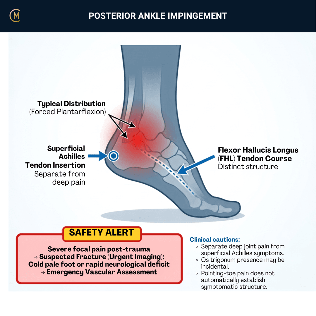
Who: Dancers, footballers, gymnasts — anyone whose sport loads end-range plantarflexion. Often a history of repetitive plantarflexion or a previous ankle sprain.
What: Compression of the tissues at the back of the ankle in forced plantarflexion.
How it behaves: Deep posterior "pinching" or ache. Worse with pointe work, kicking, jumping, push-off. Eased by avoiding plantarflexion.
The one thing to take from this page:
The os trigonum is one spoke, not the hub — and in a dancer's ankle, the scan is not the diagnosis.
It's not one thing
| What can drive it |
|---|
| Os trigonum · posterior talar process morphology · flexor hallucis longus tenosynovitis · synovitis · posterior capsule irritation · subtalar pathology |
| And chronic lateral ankle instability — which changes the odds of ending up at surgery [1] |
The imaging trap
MRI findings associated with PAIS are common in elite dancers and athletes who have no symptoms [2]. Clinical features and imaging findings correlate only partly [3].
So the diagnosis is symptom reproduction and load history — not a picture. Image to answer a question, not to confirm what your hands already told you.
⚠️ No guideline exists for this condition, anywhere. Anchored to a four-stage clinical approach from a review paper [4]. That is the closest thing to an algorithm this literature has.
2 · SE — THE SUBJECTIVE PATTERN (the 70%)
Five stages, in your course's order. The position is the diagnosis here — which makes BEHAVIOUR unusually decisive.
MAIN PROBLEM
"It pinches at the back when I go up on pointe."
"It's when I strike the ball — that follow-through position."
"There's a deep ache behind the ankle after training."
BODY CHART
- Mark it deep and posterior, between the Achilles and the peroneal tendons.
- Separate it from the Achilles itself. Achilles pain is on the tendon; this is behind and beside it.
- Note anything on the medial side with great-toe involvement — that's the FHL question.
BEHAVIOUR
| Fits posterior impingement | Points elsewhere | |
|---|---|---|
| Provoking position | Forced plantarflexion — pointe, kicking, push-off | Stretch into dorsiflexion → Achilles |
| Quality | Pinching, deep ache | Morning stiffness, thickened tendon → Achilles |
| Aggravator | The position itself | Footwear pressure → retrocalcaneal bursitis |
| Eases | Avoiding plantarflexion | — |
| Severity | VAS 0–10 at best / at worst / right now | — |
HISTORY
- Sport and volume — hours of pointe, kicking load, jump count, a recent increase.
- Previous ankle sprain, and whether the ankle has ever felt unstable since. Ask this every time (D'Hooghe et al. 2019).
- Great toe symptoms — the FHL runs right through this region and pathology there mimics and coexists with PAIS (Sharpe et al. 2020).
SPECIAL / SCREENING QUESTIONS
- Giving way, or a sense of the ankle shifting — chronic lateral instability.
- Locking or catching — an osteochondral question, not this one.
- Night or rest pain, systemic features — off this page entirely.
Assessing this region? Download the Ankle & Foot assessment template →
3 · HYPOTHESIS RANKING
| Competing pattern | The one discriminating feature |
|---|---|
| Achilles tendinopathy (mid-portion) | Morning stiffness, a thickened tendon, pain on stretch into dorsiflexion. Impingement hurts into plantarflexion, not dorsiflexion. |
| Retrocalcaneal bursitis | Tender anterior to the Achilles, with footwear pressure as the aggravator rather than the position. |
| Os trigonum / flexor hallucis longus pathology | Consider in dancers with persistent posterior pain on pointe. Image if it isn't settling — not before. |
And the one that changes the plan
Chronic lateral ankle instability, which frequently coexists. It is not a competing diagnosis so much as a modifier: it increases the likelihood of ending up at surgery in os trigonum syndrome (D'Hooghe et al. 2019), and it changes what the rehabilitation has to contain.
4 · RED FLAGS — DON'T MISS THIS
This section is free, on every pattern, forever. Safety is never gated.
| Finding | What it suggests | What you do |
|---|---|---|
| Acute trauma with inability to weight-bear or bony tenderness | Fracture | Apply the Ottawa rules. Image |
| Posterior pain after a forced plantarflexion-and-inversion injury, or persisting after one | Posterior talar process fracture — frequently missed on plain films (Majeed & McBride 2018) [17]; diagnosis was delayed in 36.4% of pooled cases (Engelmann et al. 2020) [18] | Don't file it as a sprain or as impingement. A normal x-ray doesn't clear it — ask for CT and an orthopaedic opinion |
| Locking, catching, or a recurring effusion | Osteochondral lesion | MRI, orthopaedic review |
| A hard mechanical block that doesn't change with deloading | Structural, likely os trigonum | Surgical opinion |
| Night or rest pain, systemic features, weight loss | Not this pattern | Medical referral |
| Progressive neurological symptoms into the foot | Nerve, not impingement | Medical referral |
| Marked instability or giving way | Chronic lateral instability | Reassess and address it — it changes the plan |
The rule: a posterior ankle that hurts into plantarflexion and improves when you deload it is yours. A posterior ankle that is mechanically blocked, catching, or systemically unwell is not.
Cross-links: Achilles tendinopathy · Chronic ankle instability · Lateral ankle sprain
5 · OE — THE OBJECTIVE (the 30%)
Members
Sections 5 to 7 are for members
The objective exam, management and pearls, plus the six-minute lesson and the debrief. Free with an account.
Sign in or join free6 · MX — MANAGEMENT ESSENTIALS
Members
Sections 5 to 7 are for members
The objective exam, management and pearls, plus the six-minute lesson and the debrief. Free with an account.
Sign in or join free7 · CLINICAL PEARLS + RETRIEVAL CHECK
Members
Sections 5 to 7 are for members
The objective exam, management and pearls, plus the six-minute lesson and the debrief. Free with an account.
Sign in or join freeMembers
The 4-minute lesson is for members
Twelve slides on this pattern: what you'll hear, what to examine, what to do. Free with an account.
Sign in or join freeGuideline anchor
No guideline exists for posterior ankle impingement, in Australia or anywhere.
More
Anchored to the four-stage clinical approach (Yasui et al. 2016, PMID 27795947), which is the closest thing to an algorithm in this literature, supplemented by the current diagnosis and treatment review (Ishibashi et al. 2023, PMID 36368844) and the outcome synthesis (Hamberger et al. 2025, PMID 40646513).
The anchor is a clinical algorithm from a review paper, not a guideline. There is no consensus document for this condition and no physiotherapy-specific guidance anywhere. The staged logic on this page — provoke it, deload it, rebuild capacity, refer if mechanically blocked — is drawn from that algorithm and from the athlete and dancer literature, and is labelled as such.
Members
The evidence summary is for members
Verified diagnostic accuracy, where the evidence stands and what it doesn't settle. Free with an account.
Sign in or join freeSources · 16
ShowHide
Every source on this page resolves and title-verifies against PubMed. Newest first.
- 2019D'Hooghe et al. (2019). Chronic lateral ankle instability increases the likelihood for surgery in athletes with os trigonum syndrome. Knee surgery, sports traumatology, arthroscopy : official journal of the ESSKA, 27(9), 2813-2817. https://doi.org/10.1007/s00167-018-5183-0
- 2021Baillie et al. (2021). Magnetic resonance imaging findings associated with posterior ankle impingement syndrome are prevalent in elite ballet dancers and athletes. Skeletal radiology, 50(12), 2423-2431. https://doi.org/10.1007/s00256-021-03811-x
- 2015Hayashi et al. (2015). Posterior ankle impingement in athletes: Pathogenesis, imaging features and differential diagnoses. European journal of radiology, 84(11), 2231-41. https://doi.org/10.1016/j.ejrad.2015.07.017
- 2016Yasui et al. (2016). Posterior ankle impingement syndrome: A systematic four-stage approach. World journal of orthopedics, 7(10), 657-663. https://doi.org/10.5312/wjo.v7.i10.657
- 2018Rietveld et al. (2018). Results of Treatment of Posterior Ankle Impingement Syndrome and Flexor Hallucis Longus Tendinopathy in Dancers: A Systematic Review. Journal of dance medicine & science : official publication of the International Association for Dance Medicine & Science, 22(1), 19-32. https://doi.org/10.12678/1089-313X.22.1.19
- 2020Walsh et al. (2020). Surgical Outcomes of Os Trigonum Syndrome in Dancers: A Case Series. Orthopaedic journal of sports medicine, 8(7), 2325967120938767. https://doi.org/10.1177/2325967120938767
- 2020Sharpe et al. (2020). Posterior Ankle Impingement and Flexor Hallucis Longus Pathology. Clinics in sports medicine, 39(4), 911-930. https://doi.org/10.1016/j.csm.2020.06.001
- 2022Zwiers et al. (2022). Open Versus Endoscopic Surgical Treatment of Posterior Ankle Impingement: A Meta-analysis. The American journal of sports medicine, 50(2), 563-575. https://doi.org/10.1177/03635465211004977
- 2022D'Hooghe et al. (2022). Anterior ankle impingment syndrome is less frequent, but associated with a longer absence and higher re-injury rate compared to posterior syndrome: a prospective cohort study of 6754 male professional soccer players. Knee surgery, sports traumatology, arthroscopy : official journal of the ESSKA, 30(12), 4262-4269. https://doi.org/10.1007/s00167-022-07004-4
- 2022Maffulli et al. (2022). The double posteromedial portals endoscopy for posterior ankle impingement syndrome in athletes. Journal of orthopaedics and traumatology : official journal of the Italian Society of Orthopaedics and Traumatology, 23(1), 28. https://doi.org/10.1186/s10195-022-00651-w
- 2023Ishibashi et al. (2023). Posterior Ankle Impingement Syndrome. Clinics in podiatric medicine and surgery, 40(1), 209-222. https://doi.org/10.1016/j.cpm.2022.07.014
- 2024Anastasio et al. (2024). The Utilization of Minimally Invasive Surgery for Os Trigonum Syndrome: A Systematic Review. The American journal of sports medicine, 52(8), 2168-2177. https://doi.org/10.1177/03635465231198425
- 2024Baillie et al. (2024). Single-Leg Heel Raise Capacity is Lower, and Perceived Ankle Instability is Greater, in Dancers and Athletes With Posterior Ankle Impingement Syndrome. Clinical journal of sport medicine : official journal of the Canadian Academy of Sport Medicine, 34(4), 376-380. https://doi.org/10.1097/JSM.0000000000001217
- 2024Mansur et al. (2024). Posterior Ankle Impingement: It's Not Only About the Os Trigonum. Foot & ankle orthopaedics, 9(1), 24730114241241326. https://doi.org/10.1177/24730114241241326
- 2024Gupta et al. (2024). Beyond the needle: How nonsurgical management transforms foot and ankle ability in male footballers with posterior ankle impingement- a longitudinal study. Heliyon, 10(23), e40484. https://doi.org/10.1016/j.heliyon.2024.e40484
- 2025Hamberger et al. (2025). Impingement of the ankle joint-a systematic review on the expected outcome. BMC musculoskeletal disorders, 26(1), 678. https://doi.org/10.1186/s12891-025-08785-8
Mapped to this pattern
Generic measures that also apply
- Borg RPE
Borg Rating of Perceived Exertion Scale (6-20)
perceived exertion · 1 items · 1 min
- GROC
Global Rating of Change Scale
perceived change since baseline · 1 items · 1 min
- NPRS
Numeric Pain Rating Scale
pain intensity · 1 items · 1 min
- PGIC
Patient Global Impression of Change
perceived change since baseline · 1 items · 1 min
- PSFS
Patient-Specific Functional Scale
patient-generated function · 3 items · 2 min
- SANE
Single Assessment Numeric Evaluation
patient-rated function as a percentage of normal · 1 items · 1 min
- VAS
Visual Analogue Scale
pain intensity · 1 items · 1 min
- Borg CR10
Borg Category Ratio 10 Scale
perceived exertion or symptom intensity, ratio scale · 1 items · 1 min
- GAS
Goal Attainment Scaling
individualised goal achievement · 10 min
Did this page do its job?
CliniStudy is in beta. One tap is enough — it decides what gets fixed first.