
1 · SNAPSHOT
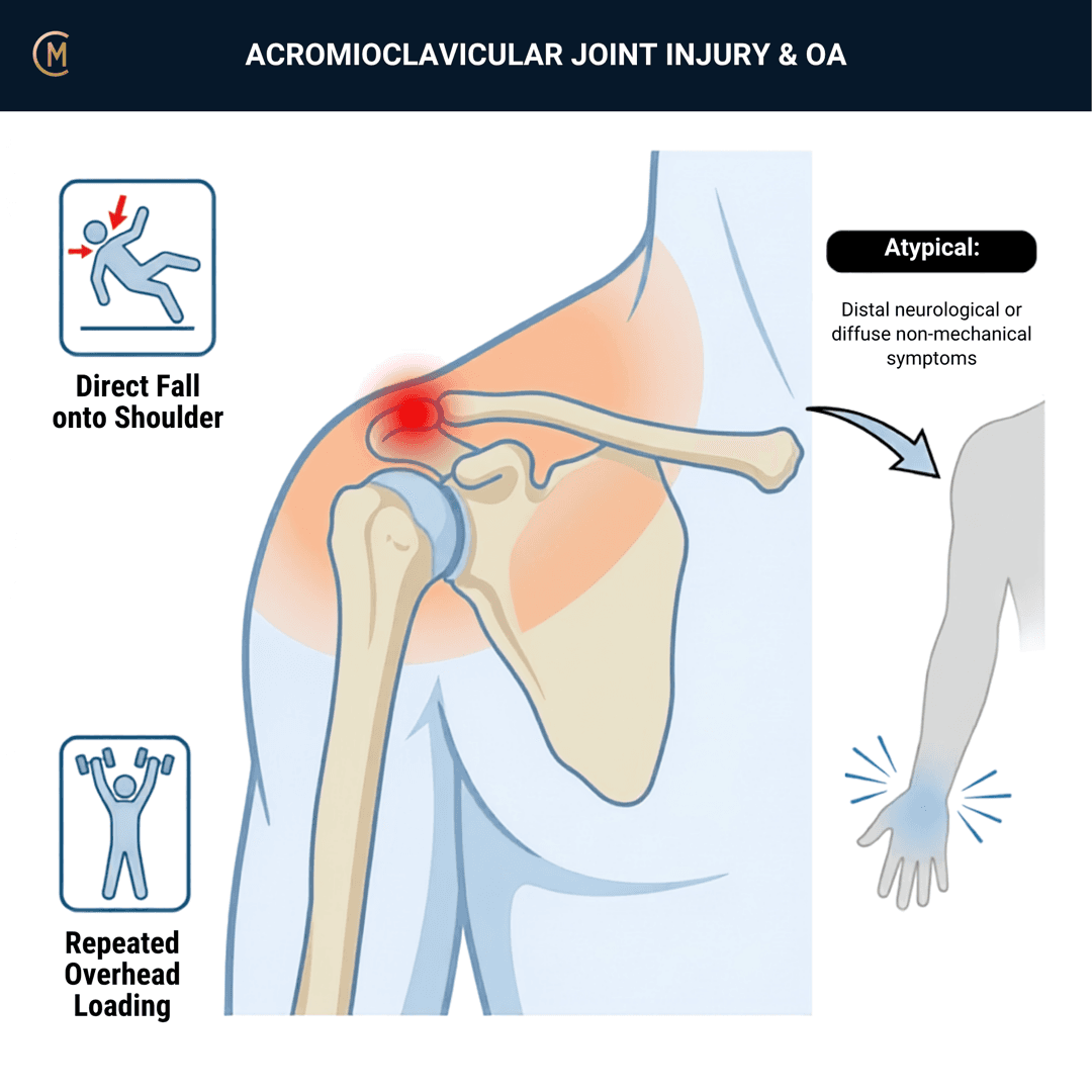
This page covers two different problems. Keep them apart in your head:
| Acute ACJ injury | ACJ osteoarthritis | |
|---|---|---|
| Onset | Fall onto the point of the shoulder, or direct contact | Gradual, repetitive load |
| Who | Young, male, sport | Older — and near-universal on imaging |
| The question | Which grade, and does it change anything | Is the joint actually the pain source |
The illness script in one line: localised pain right on the AC joint, worst with cross-body reach and overhead — from a fall onto the shoulder in a young athlete, or from nothing in particular in an older adult.
The numbers that matter:
| Figure | |
|---|---|
| Incidence, general population | 2.0 per 10,000 person-years (95% CI 1.7–2.4) |
| Incidence, young athletes | 9.2 per 1,000 person-years |
| Proportion low-grade (Rockwood I–II) in athletes | 89% |
| Proportion occurring during sport | 91% |
| Men vs women, general population | IRR 7.6 (95% CI 4.7–12.6) |
| Mean age | 39 (range 18–74) |
(General population: [1], prospective 4-year case series, n = 158. Athletes: [2], 162 injuries over 17,606 person-years.)
Days lost matters more than grade for a student's conversation: mean 10.4 days for a low-grade injury versus 63.7 days for a high-grade one (Pallis 2012).
⚠️ There is no clinical practice guideline for this pattern — Australian or international. No ACSQHC standard, no RACGP guideline, no AAOS or JOSPT CPG. Anchored to a physiotherapy-authored best-practice review [3] for conservative management of grades I–III, with the ESA–ESSKA Delphi consensus (Rosso et al. 2021) for classification. Both are older or lower-tier than the anchors elsewhere in CliniStudy, and you should say so.
Pattern > Label. The grade sets the conversation. It does not set the decision — §3.
2 · SE — THE SUBJECTIVE PATTERN (the 70%)
Two pathways, two orders. Traumatic: MAIN PROBLEM → BODY CHART → HISTORY → BEHAVIOUR. Insidious: behaviour before history.
MAIN PROBLEM
Acute: what happened, and can they lift the arm. Insidious: the task — bench press, front rack, sleeping on that side, reaching across the body.
BODY CHART
- Localised, right on the joint. They point with one finger, and that is unusual enough in the shoulder to be informative.
- May refer over the trapezius and into the anterolateral deltoid.
- No referred pain below the elbow — and that is not just a screening question here, it's one of the five variables in the only validated cluster (see below).
HISTORY — the acute pathway
- Fall onto the point of the shoulder, or direct contact. 91% happen in sport; rugby, wrestling and ice hockey have the highest rates (Pallis 2012).
- Could they keep playing? Immediate deformity? Anything visible at the time?
- Age, sex and sport: mean age 39 in the general population, and men are injured roughly 7.6 times as often (Nordin 2020).
BEHAVIOUR
- Aggs: cross-body reach, overhead, bench press and dips, carrying, lying on that side.
- Eases: arm by the side, avoiding horizontal adduction.
- 24-hr: night pain on that side is common and often the presenting complaint.
- Irritability: vigour, severity, time to settle.
The only evidence-based subjective features
Almost everything taught about the ACJ subjective pattern is convention. The exception is a cluster validated against a fluoroscopically guided anaesthetic block in 153 consecutive primary-care shoulder-pain patients [4]. Four of its five variables are history items:
| The cluster |
|---|
| Repetitive mechanism of onset |
| No referred pain below the elbow |
| No symptom provocation during passive glenohumeral abduction |
| No symptom provocation on passive external rotation |
| (the fifth: a thickened or swollen ACJ on observation) |
Two numbers worth carrying into §5: the cluster reached AUC 0.791 (95% CI 0.702–0.880), and only 22 of 153 (14%) of that primary-care population turned out to have the ACJ as their pain source at all.
SPECIAL / SCREENING QUESTIONS
General health, fever, recent infection or injection, immunosuppression, malignancy history, weight loss. See §4.
There is no study mapping ACJ pain referral from a controlled injection in healthy volunteers. The ACJ body chart you were taught is convention, not evidence. The cluster above is the part that has been tested.
Assessing this region? Download the Shoulder assessment template →
3 · HYPOTHESIS RANKING
| Competing pattern | The one discriminating feature |
|---|---|
| Rotator cuff-related pain | Lateral, not on the joint. Painful arc, painful resisted external rotation |
| Distal clavicle osteolysis | Lifter, bilateral or dominant-side, load-related. A different entity from age-related ACJ OA |
| Sternoclavicular problem | Medial end. Check it — it gets missed because nobody looks |
| Cervical referral | Symptoms below the elbow, neck movement changes it |
| Septic ACJ | Hot, swollen, systemically unwell → §4 |
The grade, and what it actually decides
Rockwood I–VI, from the source that defines all six [5]:
| Type | What is torn |
|---|---|
| I | AC ligaments sprained, joint intact |
| II | AC ligaments torn, coracoclavicular ligaments intact |
| III | Both AC and coracoclavicular torn |
| IV | Complete dislocation, posterior displacement of the clavicle into or through the trapezius fascia |
| V | Greater soft-tissue damage than III |
| VI | Inferior dislocation, subacromial or subcoracoid |
Horizontal stability comes from the AC ligaments; vertical stability from the coracoclavicular ligaments. The Zanca view is the most accurate view for the joint. The axial view is what separates type III from type IV — and an AP will not show you a posteriorly displaced clavicle.
The finding that should change how you use the grade
In 45 acute injuries read by six shoulder surgeons and six residents [6]:
| Interobserver kappa | |
|---|---|
| Agreement on the Rockwood grade | 0.52 (95% CI 0.51–0.54) |
| Agreement on the treatment chosen | 0.78 (95% CI 0.72–0.81) |
Clinicians agree on what to do more than they agree on what it is. Surgeon experience made no difference. The classification is less reliable than the decision it is supposed to drive — which is a good reason to stop treating the number as the answer.
Where the controversy actually sits: grade III. Everything below II is conservative, everything above V is usually surgical, and III is contested in every source — sub-split by some into IIIA (stable, no overriding clavicle, no scapular dysfunction → non-operative) and IIIB (overriding clavicle plus therapy-resistant scapular dysfunction → consider reconstruction). Recent reviews report a shift toward non-operative management even for type V.
Jones' hypothesis categories that change your plan: precautions (type IV, and the septic joint), sources of symptoms (ACJ vs cuff vs cervical), and patient perspectives and demand — which, per §6, drives the decision more than the grade does.
The grade sets the conversation. Four other things set the decision.
4 · RED FLAGS — DON'T MISS THIS
This section is free, on every pattern, forever.
What to actually screen
| Signs / symptoms | Implication |
|---|---|
| Hot, swollen ACJ, systemically unwell, recent injection or infection | Septic ACJ — rare and serious |
| High-energy trauma, deformity, neurovascular symptoms | Fracture · complete dislocation |
| Posterior prominence of the clavicle, skin tenting | Rockwood IV — needs the axial view |
| Skeletally immature athlete with an "ACJ injury" | Physeal injury / pseudo-dislocation — adult Rockwood reasoning does not transfer |
| Progressive weakness, symptoms below the elbow | Cervical or nerve |
| Malignancy history, weight loss, night pain unrelated to position | Malignancy |
The rare one, honestly
Septic arthritis of the ACJ exists and it is genuinely rare — only 28 cases have been reported in the entire English literature. All required a minimum of six weeks of tailored intravenous antibiotics, and two of three in one series needed open debridement. The authors say it plainly: it "requires a high index of suspicion" [7].
That is exactly what a good red flag looks like — rare enough that you will probably never see one, serious enough that you must recognise it, and honest about both.
And the one you will actually miss
Type IV. Posterior displacement of the distal clavicle into or through the trapezius fascia. It looks like a grade III on an AP. Ask for the axial view if the clavicle looks prominent posteriorly or the skin is tented (Saccomanno 2014).
Hot, swollen, systemically unwell = same-day referral, however rare it is.
Posterior prominence = ask for the axial view. An AP will not show it.
A teenager with an "ACJ separation" is usually a physeal injury.
5 · OE — THE OBJECTIVE (the 30%)
Members
Sections 5 to 7 are for members
The objective exam, management and pearls, plus the six-minute lesson and the debrief. Free with an account.
Sign in or join free6 · MX — MANAGEMENT ESSENTIALS
Members
Sections 5 to 7 are for members
The objective exam, management and pearls, plus the six-minute lesson and the debrief. Free with an account.
Sign in or join free7 · CLINICAL PEARLS + RETRIEVAL CHECK
Members
Sections 5 to 7 are for members
The objective exam, management and pearls, plus the six-minute lesson and the debrief. Free with an account.
Sign in or join freeMembers
The 4-minute lesson is for members
Twelve slides on this pattern: what you'll hear, what to examine, what to do. Free with an account.
Sign in or join freeGuideline anchor
No Australian guideline for this presentation.
More
Anchored to Acromioclavicular joint separations grades I–III: a review of the literature and development of best practice guidelines (Reid, Polson & Johnson)
There is NO clinical practice guideline for ACJ injury or ACJ OA, Australian or international. No ACSQHC standard, no RACGP guideline, no AAOS CPG, no JOSPT CPG. The anchors are a physiotherapy best-practice review and an expert Delphi — not guidelines.
Members
The evidence summary is for members
Verified diagnostic accuracy, where the evidence stands and what it doesn't settle. Free with an account.
Sign in or join freeSources · 20
ShowHide
Every source on this page resolves and title-verifies against PubMed. Newest first.
- 2020Nordin et al. (2020). Acromioclavicular joint dislocations: incidence, injury profile, and patient characteristics from a prospective case series. JSES international, 4(2), 246-250. https://doi.org/10.1016/j.jseint.2020.01.009
- 2012Pallis et al. (2012). Epidemiology of acromioclavicular joint injury in young athletes. The American journal of sports medicine, 40(9), 2072-7. https://doi.org/10.1177/0363546512450162
- 2012Reid et al. (2012). Acromioclavicular joint separations grades I-III: a review of the literature and development of best practice guidelines. Sports medicine (Auckland, N.Z.), 42(8), 681-96. https://doi.org/10.2165/11633460-000000000-00000
- 2013Cadogan et al. (2013). Shoulder pain in primary care: diagnostic accuracy of clinical examination tests for non-traumatic acromioclavicular joint pain. BMC musculoskeletal disorders, 14, 156. https://doi.org/10.1186/1471-2474-14-156
- 2014Saccomanno et al. (2014). Acromioclavicular joint instability: anatomy, biomechanics and evaluation. Joints, 2(2), 87-92. https://doi.org/10.11138/jts/2014.2.2.087
- 2023Velasquez Garcia et al. (2023). The Reliability of Acromioclavicular Joint Dislocation Classification Systems: A Comparison Between the Rockwood and Kraus Classifications. Orthopaedic journal of sports medicine, 11(2), 23259671221149391. https://doi.org/10.1177/23259671221149391
- 2020Steinmetz et al. (2020). Septic arthritis of the acromioclavicular joint: a case series and review of the literature. Shoulder & elbow, 12(4), 272-283. https://doi.org/10.1177/1758573218815289
- 2004Chronopoulos et al. (2004). Diagnostic value of physical tests for isolated chronic acromioclavicular lesions. The American journal of sports medicine, 32(3), 655-61. https://doi.org/10.1177/0363546503261723
- 2018Krill et al. (2018). A concise evidence-based physical examination for diagnosis of acromioclavicular joint pathology: a systematic review. The Physician and sportsmedicine, 46(1), 98-104. https://doi.org/10.1080/00913847.2018.1413920
- 2004Walton et al. (2004). Diagnostic values of tests for acromioclavicular joint pain. The Journal of bone and joint surgery. American volume, 86(4), 807-12. https://doi.org/10.2106/00004623-200404000-00021
- 2025Ibounig et al. (2025). Imaging abnormalities of the acromioclavicular joint and subacromial space are common in asymptomatic shoulders: a systematic review. Journal of orthopaedic surgery and research, 20(1), 7. https://doi.org/10.1186/s13018-024-05378-4
- 2022Rossano et al. (2022). Prevalence of acromioclavicular joint osteoarthritis in people not seeking care: A systematic review. Journal of orthopaedics, 32, 85-91. https://doi.org/10.1016/j.jor.2022.05.009
- 2021LeVasseur et al. (2021). Acromioclavicular Joint Injuries: Effective Rehabilitation. Open access journal of sports medicine, 12, 73-85. https://doi.org/10.2147/OAJSM.S244283
- 2015Anon. (2015). Multicenter Randomized Clinical Trial of Nonoperative Versus Operative Treatment of Acute Acromio-Clavicular Joint Dislocation. Journal of orthopaedic trauma, 29(11), 479-87. https://doi.org/10.1097/BOT.0000000000000437
- 2022Boström Windhamre et al. (2022). No difference in clinical outcome at 2-year follow-up in patients with type III and V acromioclavicular joint dislocation treated with hook plate or physiotherapy: a randomized controlled trial. Journal of shoulder and elbow surgery, 31(6), 1122-1136. https://doi.org/10.1016/j.jse.2021.12.003
- 2024Xie et al. (2024). Comparative efficacy of operative versus conservative treatment for Rockwood type III acromioclavicular joint dislocation: a systematic review and meta-analysis of randomized controlled trials. BMC musculoskeletal disorders, 25(1), 960. https://doi.org/10.1186/s12891-024-08100-x
- 2019Farrell et al. (2019). Current evidence for nonpharmacological interventions and criteria for surgical management of persistent acromioclavicular joint osteoarthritis: A systematic review. Shoulder & elbow, 11(6), 395-410. https://doi.org/10.1177/1758573219840673
- 2022Gawel et al. (2022). Criteria for return to play after operative management of acromioclavicular joint separation: a systematic review. JSES reviews, reports, and techniques, 2(2), 140-148. https://doi.org/10.1016/j.xrrt.2021.11.005
- 2016Saier et al. (2016). Return-to-activity after anatomical reconstruction of acute high-grade acromioclavicular separation. BMC musculoskeletal disorders, 17, 145. https://doi.org/10.1186/s12891-016-0989-8
- 2021Candela et al. (2021). AC joint osteoarthritis: The role of genetics. An MRI evaluation of asymptomatic elderly twins. Journal of anatomy, 238(4), 1023-1027. https://doi.org/10.1111/joa.13340
Mapped to this pattern
Generic measures that also apply
- Borg RPE
Borg Rating of Perceived Exertion Scale (6-20)
perceived exertion · 1 items · 1 min
- GROC
Global Rating of Change Scale
perceived change since baseline · 1 items · 1 min
- NPRS
Numeric Pain Rating Scale
pain intensity · 1 items · 1 min
- PGIC
Patient Global Impression of Change
perceived change since baseline · 1 items · 1 min
- PSFS
Patient-Specific Functional Scale
patient-generated function · 3 items · 2 min
- SANE
Single Assessment Numeric Evaluation
patient-rated function as a percentage of normal · 1 items · 1 min
- VAS
Visual Analogue Scale
pain intensity · 1 items · 1 min
- Borg CR10
Borg Category Ratio 10 Scale
perceived exertion or symptom intensity, ratio scale · 1 items · 1 min
- GAS
Goal Attainment Scaling
individualised goal achievement · 10 min
Did this page do its job?
CliniStudy is in beta. One tap is enough — it decides what gets fixed first.