
1 · SNAPSHOT
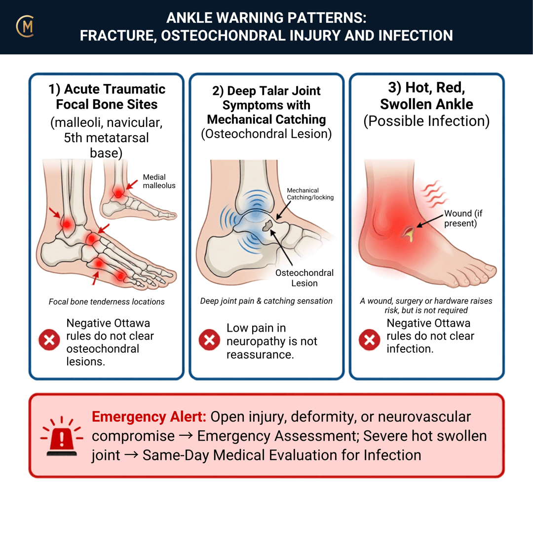
This entire page is free, permanently — all seven sections. Red flag content is never gated in CliniStudy.
Three branches, and they do not share a timeline:
| Branch | When it declares itself | What answers it |
|---|---|---|
| Fracture | Day one | The rule, then the radiograph |
| Osteochondral lesion of the talus | Week six to twelve, when the sprain won't settle | MRI, and orthopaedic review |
| Infection | Any time at all | Clinical suspicion, then urgent medical assessment |
The one thing to take from this page:
A negative Ottawa rule and a negative X-ray both clear a fracture — and nothing else.
You'll often be taught this as a day-one screen. Two of the three branches aren't.
Fracture is a day-one decision, and the Ottawa Ankle Rules are built for exactly that. The osteochondral lesion is not — it hides inside a sprain that isn't settling, and cartilage and osteochondral lesions are not rare after acute ligamentous ankle injury [1]. Infection has no timeline at all.
⚠️ No national Australian guideline governs this decision; NSW's emergency care protocol (Agency for Clinical Innovation) uses the Ottawa Ankle Rules for it. The Ottawa Ankle Rules are a decision rule about whether to X-ray, anchored to Bachmann et al. 2003 [2] and Gomes et al. 2022 [3]. Imaging referral rights differ by state and setting — check what you are permitted to request where you are working.
2 · SE — THE SUBJECTIVE PATTERN (the 70%)
Five stages — and on an acute traumatic presentation your course reorders them: MAIN PROBLEM → BODY CHART → HISTORY → BEHAVIOUR. That order is used here.
MAIN PROBLEM
"I rolled it and I heard something."
"It came up like a balloon within about twenty minutes."
"I couldn't put any weight on it at all."
And, six weeks later:
"It's still not right. There's a deep ache and it catches."
BODY CHART
- Mark the malleoli, the base of the fifth metatarsal and the navicular specifically. These are the sites the rule cares about.
- Mark swelling and bruising distribution — and note how fast it came up.
- If this is a review appointment, mark where the pain is now versus where it was. A pain that has migrated deep and central after a lateral sprain is the OLT question.
HISTORY
- Mechanism: twist, inversion, or a direct blow. Get the foot position if they can give it.
- Audible crack or pop.
- Speed of swelling. Rapid swelling and bruising within the hour is a different story from a swelling that built over a day.
- Could they weight-bear immediately, and can they take four steps now?
- Any open wound, previous surgery, or metalwork in that ankle? Fracture-related infection follows wounds, surgery, hardware and delayed healing (Krez et al. 2025).
- Diabetes, neuropathy, immunosuppression — the infection risk set.
BEHAVIOUR
- Aggs / Eases as usual — and for the OLT branch, ask specifically about deep aching after activity, catching, giving way, and swelling that keeps returning.
- Night and rest pain, fever, malaise → the infective branch.
SPECIAL / SCREENING QUESTIONS
- Fever, chills, feeling unwell — at any point in the episode, not just today.
- Is it getting better, staying the same, or getting worse? Getting worse after an acute ankle injury is not a normal trajectory.
Assessing this region? Download the Ankle & Foot assessment template →
3 · HYPOTHESIS RANKING
| Competing pattern | The one discriminating feature |
|---|---|
| Lateral ankle sprain | Ligamentous tenderness rather than bony, able to weight-bear, Ottawa rules negative. The common answer — and the one you only get to give once the others are excluded. |
| Osteochondral lesion of the talus | Deep aching with catching and a recurring effusion, persisting after a sprain that should have settled. MRI or orthopaedic review, not more loading. |
| Infection | Hot, red, swollen, with systemic symptoms — or an open wound, surgery or hardware in the history. A different pathway entirely. |
What the ranking is really doing here
On day one it is a fracture question. Apply the rule, image if it is positive, and do not label it a sprain until bony injury is excluded.
At six weeks it is a completely different question, and the ranking flips: a sprain that isn't settling makes the OLT the leading hypothesis, not the outlier. In children and adolescents this needs extra care — open physes change both the injury and the imaging (Anastasio et al. 2023), and the Ottawa rules can be applied in younger patients but with developmental and communication limits in mind (Carvalho et al. 2025).
4 · RED FLAGS — DON'T MISS THIS
This section is free, on every pattern, forever. Safety is never gated.
The escalation set
| Finding | Branch | What you do |
|---|---|---|
| Marked bony tenderness — malleoli, base of 5th MT, navicular | Fracture | Image before you treat |
| Unable to take four steps — at the time of injury or now | Fracture | Image before you treat |
| Deformity, crepitus, or an open wound | Fracture | 🚑 Emergency department |
| Hot, red, swollen + fever or malaise | Infection | 🚑 Emergency department |
| Open wound, recent surgery or hardware + escalating pain and swelling | Fracture-related infection | Same-day medical review |
| Deep pain, catching, recurring effusion at 6–12 weeks | OLT | Re-image — MRI, orthopaedic review |
| Diabetes or neuropathy + any of the above | All three | Escalate earlier. The pain signal is unreliable |
The three rules that survive everything else
1. A negative Ottawa rule clears a fracture, and nothing else. It was never designed to exclude an osteochondral lesion, a syndesmotic injury, a tendon injury or an infection.
2. A negative X-ray clears a fracture, and nothing else. It does not exclude an OLT, and it does not exclude early infection.
3. Do not call it a sprain until bony injury is excluded in anyone who fits the rule. That sentence is the failure mode.
And know the rule's limits. It is applied to an alert, cooperative patient without a significant distracting injury, intoxication, or altered sensation. Outside that population, use it if you like — but say out loud that you have.
Cross-links: Lateral ankle sprain · Syndesmotic / high ankle sprain · Infection / osteomyelitis / ulcer
5 · OE — THE OBJECTIVE (the 30%)
Members
Sections 5 to 7 are for members
The objective exam, management and pearls, plus the six-minute lesson and the debrief. Free with an account.
Sign in or join free6 · MX — MANAGEMENT ESSENTIALS
Members
Sections 5 to 7 are for members
The objective exam, management and pearls, plus the six-minute lesson and the debrief. Free with an account.
Sign in or join free7 · CLINICAL PEARLS + RETRIEVAL CHECK
Members
Sections 5 to 7 are for members
The objective exam, management and pearls, plus the six-minute lesson and the debrief. Free with an account.
Sign in or join freeMembers
The 4-minute lesson is for members
Twelve slides on this pattern: what you'll hear, what to examine, what to do. Free with an account.
Sign in or join freeGuideline anchor
No national Australian guideline governs acute ankle imaging decisions in physiotherapy; NSW's emergency care protocol (Agency for Clinical Innovation, 2023) uses the Ottawa Ankle Rules for the X-ray decision.
More
The rule's evidence is anchored to the classic systematic review (Bachmann et al. 2003, PMID 12595378) and the updated adult meta-analysis (Gomes et al. 2022, PMID 36151550). The infection limb is anchored to the IWGDF/IDSA diabetes-related foot infection guideline (Senneville et al. 2023, PMID 37779457) and, for the joint, to the septic arthritis current-concepts review (Sanpera et al. 2025, PMID 39758603). Apart from the NSW protocol, all non-Australian.
The Ottawa Ankle Rules are a decision rule about whether to X-ray, not a guideline about what a physiotherapist does next. Beyond the NSW emergency protocol there is no Australian equivalent, no Australian physiotherapy imaging-referral standard that covers every jurisdiction, and imaging referral rights differ by state and setting. Check what you are permitted to request where you are working — the page teaches the reasoning, not the paperwork.
Members
The evidence summary is for members
Verified diagnostic accuracy, where the evidence stands and what it doesn't settle. Free with an account.
Sign in or join freeSources · 19
ShowHide
Every source on this page resolves and title-verifies against PubMed. Newest first.
- 2025Baltes et al. (2025). The Prevalence, Size, and Anatomic Location of Cartilage and Osteochondral Lesions in Athletes With an Acute Ligamentous Ankle Injury. The American journal of sports medicine, 53(9), 2173-2180. https://doi.org/10.1177/03635465251344187
- 2003Bachmann et al. (2003). Accuracy of Ottawa ankle rules to exclude fractures of the ankle and mid-foot: systematic review. BMJ (Clinical research ed.), 326(7386), 417. https://doi.org/10.1136/bmj.326.7386.417
- 2022Gomes et al. (2022). Diagnostic accuracy of the Ottawa ankle rule to exclude fractures in acute ankle injuries in adults: a systematic review and meta-analysis. BMC musculoskeletal disorders, 23(1), 885. https://doi.org/10.1186/s12891-022-05831-7
- 2022Weaver et al. (2022). Magnetic resonance imaging of musculoskeletal infections. Polish journal of radiology, 87, e141-e162. https://doi.org/10.5114/pjr.2022.113825
- 2023Crowell et al. (2023). Diagnostic Imaging for Distal Extremity Injuries in Direct Access Physical Therapy: An Observational Study. International journal of sports physical therapy, 18(2), 431-438. https://doi.org/10.26603/001c.73314
- 2023Anastasio et al. (2023). Juvenile Osteochondral Lesions of the Talus: Current Concepts Review and an Update on the Literature. Children (Basel, Switzerland), 10(5). https://doi.org/10.3390/children10050884
- 2023Steele et al. (2023). Republication of "Osteochondral Lesions of the Talus: Current Concepts in Diagnosis and Treatment". Foot & ankle orthopaedics, 8(3), 24730114231192961. https://doi.org/10.1177/24730114231192961
- 2023Senneville et al. (2023). IWGDF/IDSA Guidelines on the Diagnosis and Treatment of Diabetes-related Foot Infections (IWGDF/IDSA 2023). Clinical infectious diseases : an official publication of the Infectious Diseases Society of America. https://doi.org/10.1093/cid/ciad527
- 2023Tansey et al. (2023). Current concepts in ankle fractures. Journal of clinical orthopaedics and trauma, 45, 102260. https://doi.org/10.1016/j.jcot.2023.102260
- 2024Anastasio et al. (2024). Osteochondral Lesions of the Talus: Etiology, Clinical Presentation, Treatment Options, and Outcomes. Foot and ankle clinics, 29(2), 193-211. https://doi.org/10.1016/j.fcl.2023.11.002
- 2024Maity et al. (2024). A systematic review of diabetic foot infections: pathogenesis, diagnosis, and management strategies. Frontiers in clinical diabetes and healthcare, 5, 1393309. https://doi.org/10.3389/fcdhc.2024.1393309
- 2024Zaki & Morrison (2024). Osteomyelitis and Septic Arthritis of the Foot and Ankle: Imaging Update. Clinics in podiatric medicine and surgery, 41(4), 745-758. https://doi.org/10.1016/j.cpm.2024.04.007
- 2024Butler et al. (2024). Presurgical and Postsurgical MRI Evaluation of Osteochondral Lesions of the Foot and Ankle: A Primer. Clinics in podiatric medicine and surgery, 41(4), 759-773. https://doi.org/10.1016/j.cpm.2024.04.008
- 2024Lewis et al. (2024). Rehabilitation for ankle fractures in adults. The Cochrane database of systematic reviews, 9(9), CD005595. https://doi.org/10.1002/14651858.CD005595.pub4
- 2024Cheng & Wang (2024). Advancements in the treatment of osteochondral lesions of the talus. Journal of orthopaedic surgery and research, 19(1), 827. https://doi.org/10.1186/s13018-024-05314-6
- 2025Sanpera et al. (2025). Diagnosis and management of septic arthritis: A current concepts review. Journal of children's orthopaedics, 19(1), 14-19. https://doi.org/10.1177/18632521241311302
- 2025Krez et al. (2025). Fracture-Related Infections in the Foot and Ankle. Foot and ankle clinics, 30(1), 191-200. https://doi.org/10.1016/j.fcl.2024.06.011
- 2025Carvalho et al. (2025). Ottawa Ankle Rules: A Reliable Clinical Instrument to Detect Fractures in Children and Adolescents. Revista brasileira de ortopedia, 60(1), s00441800938. https://doi.org/10.1055/s-0044-1800938
- 2025Wang & Li (2025). Early weight-bearing after ankle fracture surgery: a systematic review and meta-analysis of functional outcomes and safety. Journal of orthopaedic surgery and research, 20(1), 785. https://doi.org/10.1186/s13018-025-06216-x
Mapped to this pattern
Generic measures that also apply
- Borg RPE
Borg Rating of Perceived Exertion Scale (6-20)
perceived exertion · 1 items · 1 min
- GROC
Global Rating of Change Scale
perceived change since baseline · 1 items · 1 min
- NPRS
Numeric Pain Rating Scale
pain intensity · 1 items · 1 min
- PGIC
Patient Global Impression of Change
perceived change since baseline · 1 items · 1 min
- PSFS
Patient-Specific Functional Scale
patient-generated function · 3 items · 2 min
- SANE
Single Assessment Numeric Evaluation
patient-rated function as a percentage of normal · 1 items · 1 min
- VAS
Visual Analogue Scale
pain intensity · 1 items · 1 min
- Borg CR10
Borg Category Ratio 10 Scale
perceived exertion or symptom intensity, ratio scale · 1 items · 1 min
- GAS
Goal Attainment Scaling
individualised goal achievement · 10 min
Did this page do its job?
CliniStudy is in beta. One tap is enough — it decides what gets fixed first.