
1 · SNAPSHOT
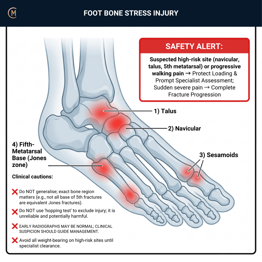
This entire page is free, permanently — all seven sections. Red flag content is never gated in CliniStudy.
Who: Runners, dancers and military personnel. Anyone after a change in training volume, intensity, surface, footwear, nutrition or recovery.
What: Repetitive loading exceeding bone remodelling capacity.
How it behaves: Gradual, focal, load-related pain with no specific trauma. Pain early in activity, and — in advanced stages — persisting afterwards. Eased by rest and unloading.
The one thing to take from this page:
The diagnosis is a load-capacity mismatch. The management is decided by WHERE.
Site decides everything
| Site | Risk | What it needs |
|---|---|---|
| Navicular | High | Image early. Often subtle and delayed. Strict offload, specialist consideration |
| Proximal 5th metatarsal (Jones) | High | Image early. Fixation is a real conversation |
| Metatarsal shafts (2nd, 3rd) | Lower | Offload, correct the load, graded return |
| Calcaneus | Lower — and it mimics plantar heel pain | Focal calcaneal tenderness + load pain → image |
Delayed diagnosis at a high-risk site buys nonunion, prolonged recovery and surgical referral [1][2].
⚠️ No Australian guideline exists. Anchored to site-specific reviews and, for the fifth metatarsal, an elite-athlete consensus.
2 · SE — THE SUBJECTIVE PATTERN (the 70%)
Five stages, in your course's order. The history is the diagnosis here — this is a load story before it is a bone story.
MAIN PROBLEM
"There's a sore spot in my foot that's getting worse the more I run."
"It used to hurt at the end of the run, now it hurts from the start."
"It's fine if I don't run."
That progression — end of activity, then during, then after — is the staging in the patient's own words.
BODY CHART
- Mark it focally. "Can you point to it with one finger?" A pain they can cover with a fingertip is a bone pain until proven otherwise.
- Name the site. Navicular, metatarsal shaft, base of the fifth, calcaneus — because the site decides the plan.
- Mark swelling if present.
BEHAVIOUR
| Fits bone stress | Points elsewhere | |
|---|---|---|
| Aggs | Weight-bearing, impact, and it's getting earlier in the session | Position and footwear → mechanical forefoot pain |
| Eases | Rest and unloading | Nothing changes it → screen wider |
| Progression | Worsening over weeks | Fluctuating with exposure → tendon or joint |
| Localisation | Focal, pointable | Diffuse, under the heads → metatarsalgia |
HISTORY — the load audit
Ask all of it, because the answer is usually in here:
- Training volume, intensity, surface and footwear — and what changed, and when.
- A return after a break. Deconditioned bone meeting old training loads.
- Recovery — sleep, rest days, back-to-back sessions.
- Nutrition and energy availability. Ask it properly.
- Previous bone stress injury. Recurrence changes the workup.
SPECIAL / SCREENING QUESTIONS
The bone-health screen — and this is not optional in a runner, an adolescent, or anyone with a recurrent or slow-healing injury:
- Energy availability, dietary restriction, weight change.
- Menstrual status where relevant. Nutrition, menstrual and bone-health variables help distinguish higher-risk injuries in female runners [3].
- Vitamin D and calcium intake. Vitamin D insufficiency is a modifiable risk factor (Knechtle et al. 2021).
- Endocrine factors, medications, prior fractures, family history.
- In adolescents: growth, nutrition, menstrual status and sport volume all matter (Beck & Drysdale 2021).
Assessing this region? Download the Ankle & Foot assessment template →
3 · HYPOTHESIS RANKING
| Competing pattern | The one discriminating feature |
|---|---|
| Lisfranc / midfoot sprain | A twist or axial load event with plantar arch bruising and pain on forefoot torsion. Bone stress is gradual with no trauma. |
| Metatarsalgia | Diffuse pain under the metatarsal heads with callus, no focal bony tenderness or percussion pain. |
| Morton's neuroma | Webspace burning radiating into the toes, with sensory change and a positive Mulder's click. |
The one that catches people out
Calcaneal stress fracture mimics plantar heel pain. Focal calcaneal tenderness plus load pain should prompt imaging rather than another six weeks of plantar fascia loading [4]. A plantar heel pain that isn't responding is a re-examination, not a re-prescription.
And know which sites you're worried about
Separate low-risk from high-risk. Navicular and proximal fifth metatarsal need earlier imaging and specialist consideration than many metatarsal shaft injuries (Paavana et al. 2024; Mandell et al. 2017).
4 · RED FLAGS — DON'T MISS THIS
This section is free, on every pattern, forever. Safety is never gated.
| Finding | What it suggests | What you do |
|---|---|---|
| Focal navicular tenderness (the "N spot") + exertional midfoot pain | High-risk navicular BSI | Image early — MRI or CT. Offload now |
| Focal tenderness at the base of the 5th metatarsal | High-risk Jones-zone injury | Image early. Offload. Specialist consideration |
| Focal bony tenderness + positive percussion or hop, with a load history | Bone stress injury | Offload and image |
| Pain now present at rest or at night | Advanced injury | Image, and stop loading |
| A normal early X-ray with a convincing story | Plain films are frequently normal early | Do not clear it. MRI |
| Recurrent bone stress injuries | Metabolic or energy-availability contribution | Metabolic workup and medical referral |
| Adolescent with midfoot pain and a nonspecific presentation | Navicular BSI presents vaguely in this group | Advanced imaging |
The three rules that survive everything else
1. Navicular and base of the fifth are high-risk. Image early. The literature is unanimous.
2. A normal early radiograph clears nothing. MRI is the preferred confirmation and grading tool. The navicular is the exception to "MRI settles it": in one athlete cohort, initial CT correlated with a correct diagnosis and initial MRI didn't always (Saxena et al. 2017) [12] — a negative MRI with a convincing N-spot story still goes back to the specialist.
3. Bone pain is a load mismatch — so check the load AND the capacity. Vitamin D and relative energy deficiency must be ruled out. Recurrent, high-risk or delayed-healing injuries should trigger a workup for vitamin D, calcium intake, endocrine factors, low energy availability and bone density risk [5].
Cross-links: Midfoot sprain (Lisfranc) · Metatarsalgia · Plantar fasciopathy
5 · OE — THE OBJECTIVE (the 30%)
Members
Sections 5 to 7 are for members
The objective exam, management and pearls, plus the six-minute lesson and the debrief. Free with an account.
Sign in or join free6 · MX — MANAGEMENT ESSENTIALS
Members
Sections 5 to 7 are for members
The objective exam, management and pearls, plus the six-minute lesson and the debrief. Free with an account.
Sign in or join free7 · CLINICAL PEARLS + RETRIEVAL CHECK
Members
Sections 5 to 7 are for members
The objective exam, management and pearls, plus the six-minute lesson and the debrief. Free with an account.
Sign in or join freeMembers
The 5-minute lesson is for members
Twelve slides on this pattern: what you'll hear, what to examine, what to do. Free with an account.
Sign in or join freeGuideline anchor
No Australian guideline covers foot bone stress injury.
More
Anchored to the foot-specific management review (Paavana et al. 2024, PMID 38435398) for the site-by-site risk stratification, the prospective MRI grading and return-to-play study (Nattiv et al. 2013, PMID 23825184) for prognosis, and the scoping review of return to running (George et al. 2024, PMID 39141251) for criteria-based progression. The elite-athlete Jones fracture consensus (Balboni et al. 2026, PMID 41451757) anchors the high-risk fifth metatarsal.
None of these is an Australian document and none is a guideline in the formal sense — the Jones fracture paper is the closest, and it is a consensus plus systematic review in elite athletes specifically. The site-by-site risk logic on this page is drawn from the primary literature and labelled as such.
Members
The evidence summary is for members
Verified diagnostic accuracy, where the evidence stands and what it doesn't settle. Free with an account.
Sign in or join freeSources · 20
ShowHide
Every source on this page resolves and title-verifies against PubMed. Newest first.
- 2024Paavana et al. (2024). Stress fractures of the foot - current evidence on management. Journal of clinical orthopaedics and trauma, 50, 102381. https://doi.org/10.1016/j.jcot.2024.102381
- 2026Modica et al. (2026). Review of Current Management of Navicular Stress Fractures in Athletes. Sports health, 18(5), 950-959. https://doi.org/10.1177/19417381251411342
- 2024Tenforde et al. (2024). Factors Associated With High-Risk and Low-Risk Bone Stress Injury in Female Runners: Implications for Risk Factor Stratification and Management. Orthopaedic journal of sports medicine, 12(5), 23259671241246227. https://doi.org/10.1177/23259671241246227
- 2018Bianchi & Luong (2018). Stress Fractures of the Calcaneus Diagnosed by Sonography: Report of 8 Cases. Journal of ultrasound in medicine : official journal of the American Institute of Ultrasound in Medicine, 37(2), 521-529. https://doi.org/10.1002/jum.14276
- 2026Nye et al. (2026). Metabolic Workup for Bone Stress Injury: A Practical Approach for Evaluating Bone Health in Athletes. Sports health, 18(3), 521-533. https://doi.org/10.1177/19417381251398501
- 2024George et al. (2024). Criteria and Guidelines for Returning to Running Following a Tibial Bone Stress Injury: A Scoping Review. Sports medicine (Auckland, N.Z.), 54(9), 2247-2265. https://doi.org/10.1007/s40279-024-02051-y
- 2013Nattiv et al. (2013). Correlation of MRI grading of bone stress injuries with clinical risk factors and return to play: a 5-year prospective study in collegiate track and field athletes. The American journal of sports medicine, 41(8), 1930-41. https://doi.org/10.1177/0363546513490645
- 2014Mayer et al. (2014). Stress fractures of the foot and ankle in athletes. Sports health, 6(6), 481-91. https://doi.org/10.1177/1941738113486588
- 2015Kahanov et al. (2015). Diagnosis, treatment, and rehabilitation of stress fractures in the lower extremity in runners. Open access journal of sports medicine, 6, 87-95. https://doi.org/10.2147/OAJSM.S39512
- 2017Shakked et al. (2017). Tarsal navicular stress fractures. Current reviews in musculoskeletal medicine, 10(1), 122-130. https://doi.org/10.1007/s12178-017-9392-9
- 2017Mandell et al. (2017). Stress fractures of the foot and ankle, part 2: site-specific etiology, imaging, and treatment, and differential diagnosis. Skeletal radiology, 46(9), 1165-1186. https://doi.org/10.1007/s00256-017-2632-7
- 2017Saxena et al. (2017). Navicular Stress Fracture Outcomes in Athletes: Analysis of 62 Injuries. The Journal of foot and ankle surgery : official publication of the American College of Foot and Ankle Surgeons, 56(5), 943-948. https://doi.org/10.1053/j.jfas.2017.06.009
- 2021Troy et al. (2021). A Narrative Review of Metatarsal Bone Stress Injury in Athletic Populations: Etiology, Biomechanics, and Management. PM & R : the journal of injury, function, and rehabilitation, 13(11), 1281-1290. https://doi.org/10.1002/pmrj.12518
- 2021Hollander et al. (2021). High union rates following surgical treatment of proximal fifth metatarsal stress fractures. Knee surgery, sports traumatology, arthroscopy : official journal of the ESSKA, 29(8), 2495-2503. https://doi.org/10.1007/s00167-021-06490-2
- 2021Knechtle et al. (2021). Vitamin D and Stress Fractures in Sport: Preventive and Therapeutic Measures-A Narrative Review. Medicina (Kaunas, Lithuania), 57(3). https://doi.org/10.3390/medicina57030223
- 2021Beck & Drysdale (2021). Risk Factors, Diagnosis and Management of Bone Stress Injuries in Adolescent Athletes: A Narrative Review. Sports (Basel, Switzerland), 9(4). https://doi.org/10.3390/sports9040052
- 2021Attia et al. (2021). Return to sport following navicular stress fracture: a systematic review and meta-analysis of three hundred and fifteen fractures. International orthopaedics, 45(10), 2699-2710. https://doi.org/10.1007/s00264-021-05147-6
- 2023Mehta et al. (2023). Tarsal Navicular Bone Stress Injuries: A Multicenter Case Series Investigating Clinical Presentation, Diagnostic Approach, Treatment, and Return to Sport in Adolescent Athletes. The American journal of sports medicine, 51(8), 2161-2168. https://doi.org/10.1177/03635465231170399
- 2024Sun et al. (2024). Risk factors of metatarsal stress fracture associated with repetitive sports activities: a systematic review. Frontiers in bioengineering and biotechnology, 12, 1435807. https://doi.org/10.3389/fbioe.2024.1435807
- 2026Balboni et al. (2026). Jones fractures in elite athletes-2025 International Foot and Ankle Sports Consensus and systematic review. Knee surgery, sports traumatology, arthroscopy : official journal of the ESSKA, 34(9), 3120-3134. https://doi.org/10.1002/ksa.70241
Mapped to this pattern
Generic measures that also apply
- Borg RPE
Borg Rating of Perceived Exertion Scale (6-20)
perceived exertion · 1 items · 1 min
- GROC
Global Rating of Change Scale
perceived change since baseline · 1 items · 1 min
- NPRS
Numeric Pain Rating Scale
pain intensity · 1 items · 1 min
- PGIC
Patient Global Impression of Change
perceived change since baseline · 1 items · 1 min
- PSFS
Patient-Specific Functional Scale
patient-generated function · 3 items · 2 min
- SANE
Single Assessment Numeric Evaluation
patient-rated function as a percentage of normal · 1 items · 1 min
- VAS
Visual Analogue Scale
pain intensity · 1 items · 1 min
- Borg CR10
Borg Category Ratio 10 Scale
perceived exertion or symptom intensity, ratio scale · 1 items · 1 min
- GAS
Goal Attainment Scaling
individualised goal achievement · 10 min
Did this page do its job?
CliniStudy is in beta. One tap is enough — it decides what gets fixed first.