
1 · SNAPSHOT
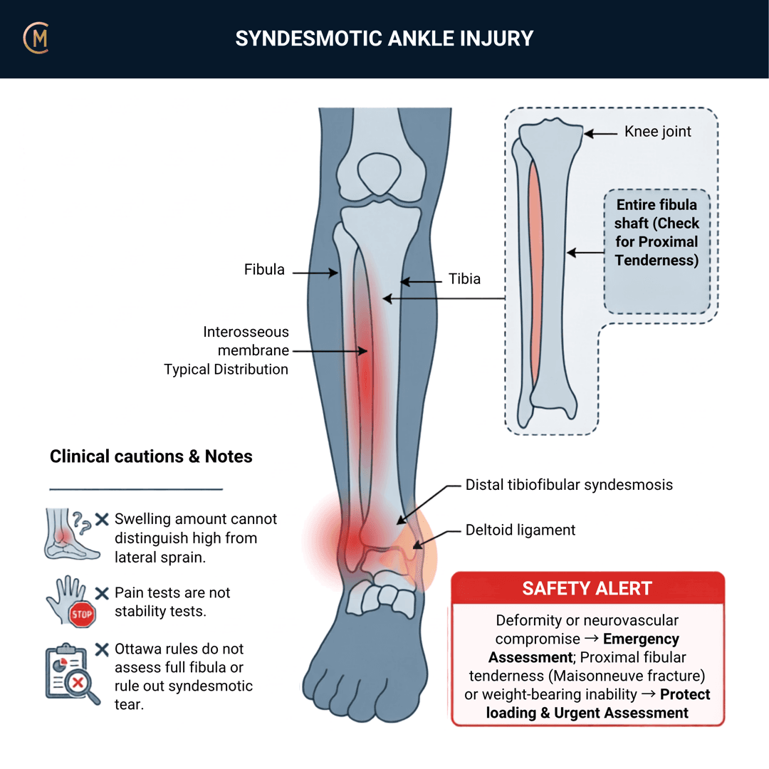
Who: Contact and cutting sports. Football, rugby, skiing, basketball, handball.
What: Pain above the joint line, after a forced dorsiflexion-and-external-rotation moment.
How it behaves: Worse on push-off, cutting and pivoting. Often less swollen than the pain suggests.
The one thing to take from this page:
It travels with the lateral sprain rather than competing with it — and the fork that matters, stable versus unstable, cannot be made clinically.
| Finding | Figure |
|---|---|
| Anterior syndesmosis injury on 3-T MRI within 10 days of an acute ligamentous ankle injury | 38% |
| Lateral ligament injury in the same 171 injuries | 73% |
| Cartilage or osteochondral lesion | 14% |
| Syndesmotic sprains as a proportion of ankle sprains, 20,336 person-years | 6.7% (4.8 per 1,000 person-years) |
| Athletics as a share of syndesmotic sprains | 81% |
([1], 166 athletes [2], US Military Academy.)
The 38% figure is already on the chronic ankle instability page. Same source, same number.
⚠️ No Australian guideline exists for syndesmotic injury. And that is verified, not asserted — an AGREE II appraisal of every ankle-sprain guideline found them only for lateral ankle ligament sprain and none for syndesmotic injury. Anchored to the ESSKA-AFAS consensus pair (2016), Level IV, whose management half rests on four non-surgical and two surgical studies.
Pattern > Label. "High ankle sprain" names a location. Stable or unstable names the decision.
2 · SE — THE SUBJECTIVE PATTERN (the 70%)
Acute and traumatic, so the order reorders: MAIN PROBLEM → BODY CHART → HISTORY → BEHAVIOUR.
MAIN PROBLEM
"I rolled my ankle" — and then, if you ask, "but it's sore up here."
BODY CHART — this is the discriminator
- Above the joint line, over the anterior tibiofibular region. Get them to point.
- Ask them to trace it upward. Pain tracking proximally along the fibula changes the question — see §4.
- Below the joint line over the ATFL is the lateral sprain. Both can be true at once.
HISTORY
- The mechanism. Forced dorsiflexion plus external rotation — a planted foot with the body rotating over it, a tackle, a ski binding, a landing.
- Contact sport, and whether they could continue.
- Previous ankle injury.
BEHAVIOUR
- Aggs: push-off, cutting, pivoting, running, stairs up.
- Eases: straight-line walking in a boot, offloading.
- Severity: VAS 0–10 at best / at worst / right now.
- Irritability: resting symptoms, vigour, time to settle.
SPECIAL / SCREENING QUESTIONS
Ability to weight-bear at the time and now, neurovascular symptoms, previous ankle surgery. See §4.
The sign worth more than any test
Pain out of proportion to the apparent injury: specificity 79%, +LR 3.05 (95% CI 1.68–5.55).
That is the only clean rule-in figure on this whole page, and it comes from 87 acute sprains across nine clinics in two Australian cities, with blinded MRI as the reference standard [3].
Note the direction. The lateral sprain's signature is swelling out of proportion to pain. This one is the reverse.
And inability to single-leg hop carries sensitivity 89% in the same cohort — a functional question you can ask before you touch them.
They rolled their ankle and it hurts more than it looks. Ask where, and ask them to trace it upward.
Assessing this region? Download the Ankle & Foot assessment template →
3 · HYPOTHESIS RANKING
| Competing pattern | The one discriminating feature |
|---|---|
| Lateral ankle sprain | At or below the joint line, swelling out of proportion to pain — see that page. And it co-occurs 38% of the time |
| Maisonneuve fracture | Proximal fibular tenderness. Outside the Ottawa zones — §4 |
| Distal fibular fracture | Ottawa positive, focal bone tenderness |
| Osteochondral lesion of the talus | Deep joint pain, catching, persistent swelling. 14% of acute injuries |
| Peroneal tendon injury | Posterolateral, resisted dorsiflexion-eversion — see the chronic ankle instability page |
| Deltoid / medial injury | Medial pain — and its presence makes instability 11× more likely |
Above the line — this is the page
Height is the discriminator, and it is free.
| Lateral sprain | Syndesmotic | |
|---|---|---|
| Location | At or below the joint line | Above it |
| Mechanism | Inversion, plantarflexion | Dorsiflexion + external rotation |
| Pain vs swelling | Swelling out of proportion to pain | Pain out of proportion to swelling |
| Ottawa rules | Apply | Apply — but they stop below the proximal fibula |
| The fork | Grade doesn't change the plan | Stable vs unstable, and it's surgical |
And they overlap. 38% of acute ligamentous ankle injuries involve the anterior syndesmosis on MRI within ten days, in a cohort where 73% had a lateral ligament injury. "Lateral OR syndesmotic" is a false choice.
One counter-intuitive finding to carry: a concomitant ATFL injury signals a different mechanism — it makes an unstable syndesmosis less likely and predicts an earlier return [4].
Jones' hypothesis categories that change your plan: sources of symptoms — and accepting there may be several — plus precautions and prognosis, which differ enormously between stable and unstable.
The question is not which one it is. It is how high it goes, and whether it is stable.
4 · RED FLAGS — DON'T MISS THIS
This section is free, on every pattern, forever.
| Signs / symptoms | Implication |
|---|---|
| Proximal fibular tenderness — palpate to the fibular head | Maisonneuve fracture — urgent assessment. The classic miss, and the Ottawa rules do not cover it |
| Ottawa Ankle Rule positive | Malleolar or midfoot fracture |
| Deep joint pain, catching, persistent swelling | Osteochondral lesion — 14% of acute ligamentous injuries |
| Unable to weight-bear, gross instability, deformity | Unstable syndesmosis or fracture-dislocation — emergency department if deformed or neurovascular change; otherwise prompt medical review |
| Escalating pain, tense compartment, neurological symptoms | Compartment syndrome — emergency |
| Calf pain, swelling, warmth after immobilisation | DVT |
The Ottawa rules stop below the fibular head
Ottawa Ankle Rules: pooled negative likelihood ratio 0.08 (95% CI 0.03–0.18) across 27 studies and 15,581 patients [5]. Excellent — for what it covers.
It covers the malleolar and midfoot zones only. A negative Ottawa rule says nothing about a Maisonneuve fracture of the proximal fibula.
So palpate the whole fibula on every ankle sprain — not only the high-energy ones. (No verified incidence figure for Maisonneuve fracture exists.)
And plain films miss the syndesmosis itself
| Radiograph detects a syndesmotic injury | about half on the anteroposterior view, two-thirds on the mortise view |
| Intra-operative stress radiography | missed about half of instabilities confirmed at arthroscopy |
[6]
Why missing it matters
Unstable syndesmotic lesions are always managed by surgery, and untreated instability leads to persistent pain, chronic instability and ankle osteoarthritis [7].
That is the whole argument for slowing down on this one. A missed lateral sprain grade is a nuisance. A missed unstable syndesmosis is a joint.
Palpate to the fibular head on every ankle sprain. The Ottawa rules stop below it.
A normal X-ray does not clear the syndesmosis — it misses about half of them.
Untreated instability ends in osteoarthritis. That is why this pattern is amber.
5 · OE — SYNDESMOSIS AND ASSOCIATED-INJURY ASSESSMENT
Members
Sections 5 to 7 are for members
The objective exam, management and pearls, plus the six-minute lesson and the debrief. Free with an account.
Sign in or join free6 · MX — MANAGEMENT ESSENTIALS
Members
Sections 5 to 7 are for members
The objective exam, management and pearls, plus the six-minute lesson and the debrief. Free with an account.
Sign in or join free7 · CLINICAL PEARLS + RETRIEVAL CHECK
Members
Sections 5 to 7 are for members
The objective exam, management and pearls, plus the six-minute lesson and the debrief. Free with an account.
Sign in or join freeMembers
The 5-minute lesson is for members
Twelve slides on this pattern: what you'll hear, what to examine, what to do. Free with an account.
Sign in or join freeGuideline anchor
No Australian guideline for this presentation.
More
Anchored to ESSKA-AFAS consensus and guidelines on acute isolated syndesmotic injury — classification and diagnosis (van Dijk et al., PMID 26704800) paired with conservative and surgical management (PMID 26846658)
An AGREE II appraisal of every ankle-sprain guideline found them only for LATERAL ankle ligament sprain — Netherlands, USA, Canada and Australia (nursing) — and NONE for syndesmotic injury (Green et al. 2019, PMID 31470826). But the clinical accuracy data on this page IS Australian: Sman (University of Sydney, nine clinics) and Alonso (Bankstown NSW physiotherapists).
Members
The evidence summary is for members
Verified diagnostic accuracy, where the evidence stands and what it doesn't settle. Free with an account.
Sign in or join freeSources · 26
ShowHide
Every source on this page resolves and title-verifies against PubMed. Newest first.
- 2025Baltes et al. (2025). The Prevalence, Size, and Anatomic Location of Cartilage and Osteochondral Lesions in Athletes With an Acute Ligamentous Ankle Injury. The American journal of sports medicine, 53(9), 2173-2180. https://doi.org/10.1177/03635465251344187
- 2011Waterman et al. (2011). Risk factors for syndesmotic and medial ankle sprain: role of sex, sport, and level of competition. The American journal of sports medicine, 39(5), 992-8. https://doi.org/10.1177/0363546510391462
- 2015Sman et al. (2015). Diagnostic accuracy of clinical tests for ankle syndesmosis injury. British journal of sports medicine, 49(5), 323-9. https://doi.org/10.1136/bjsports-2013-092787
- 2016Calder et al. (2016). Stable Versus Unstable Grade II High Ankle Sprains: A Prospective Study Predicting the Need for Surgical Stabilization and Time to Return to Sports. Arthroscopy : the journal of arthroscopic & related surgery : official publication of the Arthroscopy Association of North America and the International Arthroscopy Association, 32(4), 634-42. https://doi.org/10.1016/j.arthro.2015.10.003
- 2003Bachmann et al. (2003). Accuracy of Ottawa ankle rules to exclude fractures of the ankle and mid-foot: systematic review. BMJ (Clinical research ed.), 326(7386), 417. https://doi.org/10.1136/bmj.326.7386.417
- 2018Kellett et al. (2018). Diagnostic imaging of ankle syndesmosis injuries: A general review. Journal of medical imaging and radiation oncology, 62(2), 159-168. https://doi.org/10.1111/1754-9485.12708
- 2021Corte-Real & Caetano (2021). Ankle and syndesmosis instability: consensus and controversies. EFORT open reviews, 6(6), 420-431. https://doi.org/10.1302/2058-5241.6.210017
- 2006Lin et al. (2006). Ankle syndesmosis injuries: anatomy, biomechanics, mechanism of injury, and clinical guidelines for diagnosis and intervention. The Journal of orthopaedic and sports physical therapy, 36(6), 372-84. https://doi.org/10.2519/jospt.2006.2195
- 2016van Dijk et al. (2016). Classification and diagnosis of acute isolated syndesmotic injuries: ESSKA-AFAS consensus and guidelines. Knee surgery, sports traumatology, arthroscopy : official journal of the ESSKA, 24(4), 1200-16. https://doi.org/10.1007/s00167-015-3942-8
- 2016van Dijk et al. (2016). Conservative and surgical management of acute isolated syndesmotic injuries: ESSKA-AFAS consensus and guidelines. Knee surgery, sports traumatology, arthroscopy : official journal of the ESSKA, 24(4), 1217-27. https://doi.org/10.1007/s00167-016-4017-1
- 2019Green et al. (2019). What is the quality of clinical practice guidelines for the treatment of acute lateral ankle ligament sprains in adults? A systematic review. BMC musculoskeletal disorders, 20(1), 394. https://doi.org/10.1186/s12891-019-2750-6
- 2013Sman et al. (2013). Diagnostic accuracy of clinical tests for diagnosis of ankle syndesmosis injury: a systematic review. British journal of sports medicine, 47(10), 620-8. https://doi.org/10.1136/bjsports-2012-091702
- 2021Netterström-Wedin & Bleakley (2021). Diagnostic accuracy of clinical tests assessing ligamentous injury of the ankle syndesmosis: A systematic review with meta-analysis. Physical therapy in sport : official journal of the Association of Chartered Physiotherapists in Sports Medicine, 49, 214-226. https://doi.org/10.1016/j.ptsp.2021.03.005
- 1998Alonso et al. (1998). Clinical tests for ankle syndesmosis injury: reliability and prediction of return to function. The Journal of orthopaedic and sports physical therapy, 27(4), 276-84. https://doi.org/10.2519/jospt.1998.27.4.276
- 2011Kellett (2011). The clinical features of ankle syndesmosis injuries: a general review. Clinical journal of sport medicine : official journal of the Canadian Academy of Sport Medicine, 21(6), 524-9. https://doi.org/10.1097/JSM.0b013e318234be7d
- 2022Spindler et al. (2022). A systematic review and meta-analysis on the value of the external rotation stress test under fluoroscopy to detect syndesmotic injuries. EFORT open reviews, 7(10), 671-679. https://doi.org/10.1530/EOR-22-0037
- 2022Ng et al. (2022). Which test is the best? An updated literature review of imaging modalities for acute ankle diastasis injuries. Journal of medical radiation sciences, 69(3), 382-393. https://doi.org/10.1002/jmrs.589
- 2022Ashkani Esfahani et al. (2022). Volume measurements on weightbearing computed tomography can detect subtle syndesmotic instability. Journal of orthopaedic research : official publication of the Orthopaedic Research Society, 40(2), 460-467. https://doi.org/10.1002/jor.25049
- 2022Raheman et al. (2022). Can Weightbearing Cone-beam CT Reliably Differentiate Between Stable and Unstable Syndesmotic Ankle Injuries? A Systematic Review and Meta-analysis. Clinical orthopaedics and related research, 480(8), 1547-1562. https://doi.org/10.1097/CORR.0000000000002171
- 2019Hagemeijer et al. (2019). Range of Normal and Abnormal Syndesmotic Measurements Using Weightbearing CT. Foot & ankle international, 40(12), 1430-1437. https://doi.org/10.1177/1071100719866831
- 2003Oae et al. (2003). Injury of the tibiofibular syndesmosis: value of MR imaging for diagnosis. Radiology, 227(1), 155-61. https://doi.org/10.1148/radiol.2271011865
- 2023Bolia et al. (2023). Elite Athletes Successfully Return to the Preinjury Level of Sport Following Ankle Syndesmosis Injuries: A Systematic Review and Meta-Analysis. Clinical journal of sport medicine : official journal of the Canadian Academy of Sport Medicine, 33(1), 90-96. https://doi.org/10.1097/JSM.0000000000001019
- 2022Salameh et al. (2022). Return to Play After Isolated Syndesmotic Ligamentous Injury in Athletes: A Systematic Review and Meta-analysis. Foot & ankle orthopaedics, 7(2), 24730114221096482. https://doi.org/10.1177/24730114221096482
- 2019Vancolen et al. (2019). Return to Sport After Ankle Syndesmotic Injury: A Systematic Review. Sports health, 11(2), 116-122. https://doi.org/10.1177/1941738118816282
- 2020D'Hooghe et al. (2020). Return to play after surgery for isolated unstable syndesmotic ankle injuries (West Point grade IIB and III) in 110 male professional football players: a retrospective cohort study. British journal of sports medicine, 54(19), 1168-1173. https://doi.org/10.1136/bjsports-2018-100298
- 2012Howard et al. (2012). Magnetic resonance imaging as a predictor of return to play following syndesmosis (high) ankle sprains in professional football players. Sports health, 4(6), 535-43. https://doi.org/10.1177/1941738112462531
Mapped to this pattern
Generic measures that also apply
- Borg RPE
Borg Rating of Perceived Exertion Scale (6-20)
perceived exertion · 1 items · 1 min
- GROC
Global Rating of Change Scale
perceived change since baseline · 1 items · 1 min
- NPRS
Numeric Pain Rating Scale
pain intensity · 1 items · 1 min
- PGIC
Patient Global Impression of Change
perceived change since baseline · 1 items · 1 min
- PSFS
Patient-Specific Functional Scale
patient-generated function · 3 items · 2 min
- SANE
Single Assessment Numeric Evaluation
patient-rated function as a percentage of normal · 1 items · 1 min
- VAS
Visual Analogue Scale
pain intensity · 1 items · 1 min
- Borg CR10
Borg Category Ratio 10 Scale
perceived exertion or symptom intensity, ratio scale · 1 items · 1 min
- GAS
Goal Attainment Scaling
individualised goal achievement · 10 min
Did this page do its job?
CliniStudy is in beta. One tap is enough — it decides what gets fixed first.