
1 · SNAPSHOT
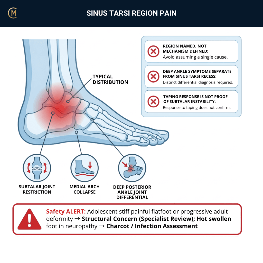
The sprain that never finished, and the reason a "simple" lateral ankle keeps coming back.
Illness script in one line: deep, poorly localised pain in the hollow just in front of and below the lateral malleolus, weeks to months after an ankle sprain, with a sense of giving way on uneven ground rather than a clear painful movement.
Caseload frequency: uncommon as a referral label, common as the reason an ankle sprain is still on your list at week ten.
A sinus tarsi is a place, not a diagnosis
This is the organising idea. The tarsal sinus is an anatomical space — a funnel between the talus and calcaneus holding the interosseous talocalcaneal ligament, fat, vessels and nerve endings — and there are three different ways to end up with symptoms in it.
| Route in | What you'd expect alongside it |
|---|---|
| 1. The sprain that never finished | deep lateral ankle pain post-sprain, instability or giving way on uneven ground. Lateral ligament laxity, poor balance, a history of repeat sprains |
| 2. The collapsing medial column | A flattening arch, medial hindfoot symptoms, too-many-toes. Posterior tibial tendon injury has been associated with spring ligament injury and sinus tarsi abnormality [1] and posterior tibial tendon abnormalities associated with abnormal signal intensity in the sinus tarsi [2]. |
| 3. Something else in the joint | An accessory ossicle has been reported in possible sinus tarsi syndrome [3]. Coalition, osteochondral lesion, occult fracture, arthritis. Section 4 |
Two radiology papers link this space to the posterior tibial tendon. That is why the medial column is on this page at all, and it is the thing a student will not expect.
And the honest headline
A systematic review has concluded that currently used imaging options cannot accurately predict subtalar joint instability [4].
Read that twice. It means the scan will not settle this for you, and your examination and your reasoning are what the diagnosis rests on.
2 · SE — THE SUBJECTIVE PATTERN (the 70%)
Main problem
"It aches deep in there and I don't trust it on uneven ground." deep lateral ankle pain post-sprain. Instability or "giving way" on uneven ground.
Two features to listen for, and they matter more than any test on this page: the pain is deep and hard to localise, and the instability is terrain-dependent rather than movement-dependent.
Body chart
Ask them to point, and expect a fingertip going into the hollow anterior and inferior to the lateral malleolus rather than onto a ligament.
Then ask them to point on the other side of the foot too. A patient who also puts a hand along the medial arch or behind the medial malleolus is telling you about route two.
Behaviour
Aggs: uneven ground, grass, sand, gravel, trail; walking on a camber; prolonged standing and walking; first steps after rest; turning on the foot.
Eases: flat, predictable surfaces; supportive footwear; taping or a brace; rest.
Severity: VAS 0–10 at best / at worst / right now.
"Uneven ground" is close to the defining aggravating factor. A subtalar joint's job is to accommodate the ground, so a subtalar problem shows up when the ground stops being flat — and that is a far more useful question than asking about inversion.
History
- The sprain. When, how bad, how it was managed, and — the key question — did it ever fully settle?
- How many sprains? Repeat sprains change the conversation.
- Or no sprain at all? Then think harder about routes two and three — a gradually flattening foot, or a bony cause. Section 4.
- Footwear, work surfaces, sport surfaces.
- Any change in foot shape? Ask directly, and ask whether anyone has mentioned flat feet.
Special / screening questions
- Any true locking, catching, or an inability to weight-bear? Section 4.
- Night pain, or pain unrelated to activity? Section 4.
- Adolescent, with a stiff and painful flat foot? Section 4.
- Progressive flattening of the arch, particularly in an adult over 40? Section 4.
Assessing this region? Download the Ankle & Foot assessment template →
3 · HYPOTHESIS RANKING
The chronic lateral ankle complex differential sets the field.
| Pattern | The one discriminating feature |
|---|---|
| Sinus tarsi syndrome | Deep, poorly localised pain in the tarsal sinus, tender on direct pressure into the hollow, worse on uneven ground, after a sprain. |
| Chronic ankle instability (lateral ligament) | Discrete ATFL tenderness, a positive anterior drawer, and giving way on a specific movement rather than on terrain. Its own page — and differences between subtalar and lateral ankle instability have been examined specifically [5]. They overlap; they are not identical. |
| Subtalar instability | Overlaps heavily with this page, and imaging cannot settle it (Krähenbühl et al. 2019). Often the same patient. |
| Posterior tibial tendon dysfunction / progressive collapsing foot | Medial symptoms, a flattening arch, too-many-toes, a poor single-leg heel raise. Associated with sinus tarsi abnormality in two separate papers (Balen et al. 2001; Anderson et al. 2000). Route two. |
| Osteochondral lesion of the talus | Deep pain, catching, swelling, and an ankle that never settled after a sprain. Section 4. |
| Tarsal coalition | An adolescent, a stiff and painful flat foot, recurrent "sprains", limited subtalar motion. Section 4. |
| Accessory ossicle or occult fracture | Reported in possible sinus tarsi syndrome (Achong et al. 2020). Section 4. |
The discriminator that does most of the work: is subtalar motion free, and is the medial column holding up?
4 · RED FLAGS — DON'T MISS THIS
Free forever. Never gated.
1. The ankle that never settled after a sprain
This is the one, and it is the reason patients arrive on this page.
An ankle that has not improved in the expected timeframe after a sprain has a differential of its own: an osteochondral lesion of the talus, an occult fracture, an accessory ossicle, or a persistent syndesmotic or subtalar problem. An unsuspected accessory ossicle has been reported in possible sinus tarsi syndrome [3] and sinus tarsi syndrome reported as diagnosed on bone SPECT/CT [6]. Add a talar process fracture to that list: lateral process ("snowboarder's") fractures come from forced dorsiflexion and inversion and are frequently missed on plain X-ray (Majeed & McBride 2018) [21]. High clinical suspicion with a normal film → CT is the next question, not reassurance.
Escalate on: true catching or locking, an inability to weight-bear, night pain, focal bony tenderness, or a persistent effusion. "It's been three months and it hasn't changed" is itself the trigger.
2. The adolescent with a stiff, painful flat foot
Recurrent ankle "sprains" in an adolescent with limited subtalar motion and a rigid flat foot is a tarsal coalition until someone proves otherwise. This needs imaging and an opinion, not balance retraining — and the giveaway is that subtalar inversion and eversion are restricted rather than merely painful.
3. The adult whose arch is collapsing
Posterior tibial tendon injury has been associated with spring ligament injury and sinus tarsi abnormality [1] and posterior tibial tendon abnormalities associated with abnormal signal intensity in the sinus tarsi [2].
A progressive collapsing foot in an adult — a flattening arch, a too-many-toes sign, an inability to perform a single-leg heel raise — is a different problem with a different urgency, and it is progressive. Balance exercises for a collapsing medial column is the specific error this page exists to prevent.
4. The hot, swollen or systemically unwell foot
A hot, swollen, exquisitely tender hindfoot without a mechanical story is an infection, crystal or inflammatory question and it goes today.
5 · OE — THE OBJECTIVE (the 30%)
Members
Sections 5 to 7 are for members
The objective exam, management and pearls, plus the six-minute lesson and the debrief. Free with an account.
Sign in or join free6 · MX — MANAGEMENT ESSENTIALS
Members
Sections 5 to 7 are for members
The objective exam, management and pearls, plus the six-minute lesson and the debrief. Free with an account.
Sign in or join free7 · CLINICAL PEARLS + RETRIEVAL CHECK
Members
Sections 5 to 7 are for members
The objective exam, management and pearls, plus the six-minute lesson and the debrief. Free with an account.
Sign in or join freeMembers
The 5-minute lesson is for members
Twelve slides on this pattern: what you'll hear, what to examine, what to do. Free with an account.
Sign in or join freeGuideline anchor
No Australian guideline and no clinical practice guideline of any kind.
More
The clinical anchors are reviews of subtalar instability — its diagnosis and treatment (Barg et al. 2012, PMID 22381348), its aetiology, diagnosis and management (Keefe et al. 2002, PMID 12512411) and recent developments in the treatment of ankle and subtalar instability (Sugimoto et al. 2017, PMID 28979582). The most important single finding in our library is a systematic review concluding that currently used imaging options cannot accurately predict subtalar joint instability (Krähenbühl et al. 2019, PMID 30367197). The physiotherapy-facing anchor is an Archives of Physical Medicine and Rehabilitation paper on the importance of biomechanically-based evaluation and treatment (Shear et al. 1993, PMID 8328905).
None of these is a clinical practice guideline. They are narrative reviews, one systematic review of imaging, imaging series, surgical case series and one older rehabilitation paper. There is no modern physiotherapy trial for this condition in our library.
Members
The evidence summary is for members
Verified diagnostic accuracy, where the evidence stands and what it doesn't settle. Free with an account.
Sign in or join freeSources · 20
ShowHide
Every source on this page resolves and title-verifies against PubMed. Newest first.
- 2001Balen & Helms (2001). Association of posterior tibial tendon injury with spring ligament injury, sinus tarsi abnormality, and plantar fasciitis on MR imaging. AJR. American journal of roentgenology, 176(5), 1137-43. https://doi.org/10.2214/ajr.176.5.1761137
- 2000Anderson et al. (2000). Association of posterior tibial tendon abnormalities with abnormal signal intensity in the sinus tarsi on MR imaging. Skeletal radiology, 29(9), 514-9. https://doi.org/10.1007/s002560000248
- 2020Achong (2020). Unsuspected Accessory Ossicle in Possible Sinus Tarsi Syndrome: The Value of Bone SPECT/CT. Clinical nuclear medicine, 45(4), e215-e216. https://doi.org/10.1097/RLU.0000000000002943
- 2019Krähenbühl et al. (2019). Currently used imaging options cannot accurately predict subtalar joint instability. Knee surgery, sports traumatology, arthroscopy : official journal of the ESSKA, 27(9), 2818-2830. https://doi.org/10.1007/s00167-018-5232-8
- 2018Yoon et al. (2018). Differences Between Subtalar Instability and Lateral Ankle Instability Focusing on Subtalar Ligaments Based on Three Dimensional Isotropic Magnetic Resonance Imaging. Journal of computer assisted tomography, 42(4), 566-573. https://doi.org/10.1097/RCT.0000000000000717
- 2020Mahato et al. (2020). Sinus Tarsi Syndrome: Diagnosed on (99m)Tc-MDP bone SPECT/CT. Asia Oceania journal of nuclear medicine & biology, 8(2), 153-156. https://doi.org/10.22038/AOJNMB.2020.45897.1309
- 2017Kim et al. (2017). Subtalar instability: imaging features of subtalar ligaments on 3D isotropic ankle MRI. BMC musculoskeletal disorders, 18(1), 475. https://doi.org/10.1186/s12891-017-1841-5
- 2001Lektrakul et al. (2001). Tarsal sinus: arthrographic, MR imaging, MR arthrographic, and pathologic findings in cadavers and retrospective study data in patients with sinus tarsi syndrome. Radiology, 219(3), 802-10. https://doi.org/10.1148/radiology.219.3.r01jn31802
- 1993Klein & Spreitzer (1993). MR imaging of the tarsal sinus and canal: normal anatomy, pathologic findings, and features of the sinus tarsi syndrome. Radiology, 186(1), 233-40. https://doi.org/10.1148/radiology.186.1.8416571
- 1990Beltran et al. (1990). Ligaments of the lateral aspect of the ankle and sinus tarsi: an MR imaging study. Radiology, 177(2), 455-8. https://doi.org/10.1148/radiology.177.2.2217784
- 2016Lee et al. (2016). Diagnostic validity of alternative manual stress radiographic technique detecting subtalar instability with concomitant ankle instability. Knee surgery, sports traumatology, arthroscopy : official journal of the ESSKA, 24(4), 1029-39. https://doi.org/10.1007/s00167-016-4037-x
- 1993Shear et al. (1993). Sinus tarsi syndrome: the importance of biomechanically-based evaluation and treatment. Archives of physical medicine and rehabilitation, 74(7), 777-81. https://doi.org/10.1016/0003-9993(93)90044-b
- 1994Kuwada (1994). Long-term retrospective analysis of the treatment of sinus tarsi syndrome. The Journal of foot and ankle surgery : official publication of the American College of Foot and Ankle Surgeons, 33(1), 28-9. PubMed 8161989
- 2019Mansur et al. (2019). Subtalar arthroscopic debridment for the treatment of sinus tarsi syndrome: case series. Revista da Associacao Medica Brasileira (1992), 65(3), 370-374. https://doi.org/10.1590/1806-9282.65.3.370
- 2018Li et al. (2018). Arthroscopic treatment combined with the ankle stabilization procedure is effective for sinus tarsi syndrome in patients with chronic ankle instability. Knee surgery, sports traumatology, arthroscopy : official journal of the ESSKA, 26(10), 3135-3139. https://doi.org/10.1007/s00167-017-4813-2
- 1998Karlsson et al. (1998). Subtalar instability of the foot. A review and results after surgical treatment. Scandinavian journal of medicine & science in sports, 8(4), 191-7. https://doi.org/10.1111/j.1600-0838.1998.tb00191.x
- 1997Thermann et al. (1997). Treatment algorithm of chronic ankle and subtalar instability. Foot & ankle international, 18(3), 163-9. https://doi.org/10.1177/107110079701800309
- 2017Sugimoto et al. (2017). Recent Developments in the Treatment of Ankle and Subtalar Instability. The open orthopaedics journal, 11, 687-696. https://doi.org/10.2174/1874325001711010687
- 2012Barg et al. (2012). Subtalar instability: diagnosis and treatment. Foot & ankle international, 33(2), 151-60. https://doi.org/10.3113/FAI.2012.0151
- 2002Keefe & Haddad (2002). Subtalar instability. Etiology, diagnosis, and management. Foot and ankle clinics, 7(3), 577-609. https://doi.org/10.1016/s1083-7515(02)00047-5
Mapped to this pattern
Generic measures that also apply
- Borg RPE
Borg Rating of Perceived Exertion Scale (6-20)
perceived exertion · 1 items · 1 min
- GROC
Global Rating of Change Scale
perceived change since baseline · 1 items · 1 min
- NPRS
Numeric Pain Rating Scale
pain intensity · 1 items · 1 min
- PGIC
Patient Global Impression of Change
perceived change since baseline · 1 items · 1 min
- PSFS
Patient-Specific Functional Scale
patient-generated function · 3 items · 2 min
- SANE
Single Assessment Numeric Evaluation
patient-rated function as a percentage of normal · 1 items · 1 min
- VAS
Visual Analogue Scale
pain intensity · 1 items · 1 min
- Borg CR10
Borg Category Ratio 10 Scale
perceived exertion or symptom intensity, ratio scale · 1 items · 1 min
- GAS
Goal Attainment Scaling
individualised goal achievement · 10 min
Did this page do its job?
CliniStudy is in beta. One tap is enough — it decides what gets fixed first.